

Prolonged helium postconditioning protocols during early reperfusion do not induce cardioprotection in the rat heart in vivo: role of inflammatory cytokines
- PMID: 25759838
- PMCID: PMC4352470
- DOI: 10.1155/2015/216798
Prolonged helium postconditioning protocols during early reperfusion do not induce cardioprotection in the rat heart in vivo: role of inflammatory cytokines
Abstract
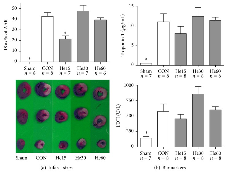
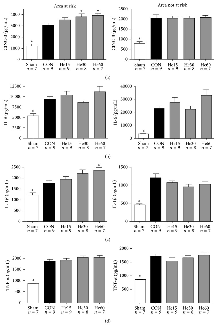
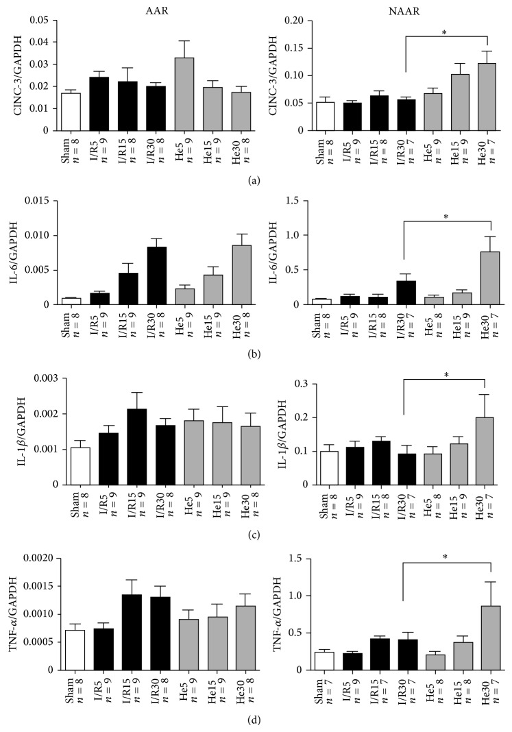
Postconditioning of myocardial tissue employs short cycles of ischemia or pharmacologic agents during early reperfusion. Effects of helium postconditioning protocols on infarct size and the ischemia/reperfusion-induced immune response were investigated by measurement of protein and mRNA levels of proinflammatory cytokines. Rats were anesthetized with S-ketamine (150 mg/kg) and diazepam (1.5 mg/kg). Regional myocardial ischemia/reperfusion was induced; additional groups inhaled 15, 30, or 60 min of 70% helium during reperfusion. Fifteen minutes of helium reduced infarct size from 43% in control to 21%, whereas 30 and 60 minutes of helium inhalation led to an infarct size of 47% and 39%, respectively. Increased protein levels of cytokine-induced neutrophil chemoattractant (CINC-3) and interleukin-1 beta (IL-1β) were found after 30 or 60 min of helium inhalation, in comparison to control. 30 min of helium increased mRNA levels of CINC-3, IL-1β, interleukin 6 (IL-6), and tumor necrosis factor alpha (TNF-α) in myocardial tissue not directly subjected to ischemia/reperfusion. These results suggest that the effectiveness of the helium postconditioning protocol is very sensitive to duration of noble gas application. Additionally, helium was associated with higher levels of inflammatory cytokines; however, it is not clear whether this is causative of nature or part of an epiphenomenon.
Figures
References
-
- Ovize M., Baxter G. F., di Lisa F., et al. Postconditioning and protection from reperfusion injury: where do we stand: position paper from the Working Group of Cellular Biology of the Heart of the European Society of Cardiology. Cardiovascular Research. 2010;87(3):406–423. doi: 10.1093/cvr/cvq129. - DOI - PubMed
-
- Girn H. R., Ahilathirunayagam S., Mavor A. I., Homer-Vanniasinkam S. Reperfusion syndrome: cellular mechanisms of microvascular dysfunction and potential therapeutic strategies. Vascular and Endovascular Surgery. 2007;41(4):277–293. - PubMed
-
- Zhao Z. Q., Corvera J. S., Halkos M. E., et al. Inhibition of myocardial injury by ischemic postconditioning during reperfusion: comparison with ischemic preconditioning. American Journal of Physiology. Heart and Circulatory Physiology. 2003;285(2):H579–H588. doi: 10.1152/ajpheart.01064.2002. - DOI - PubMed
MeSH terms
Substances
LinkOut - more resources
Full Text Sources
Other Literature Sources
