

Crystal Structure of Human Profilaggrin S100 Domain and Identification of Target Proteins Annexin II, Stratifin, and HSP27
- PMID: 25760235
- PMCID: PMC4466033
- DOI: 10.1038/jid.2015.102
Crystal Structure of Human Profilaggrin S100 Domain and Identification of Target Proteins Annexin II, Stratifin, and HSP27
Abstract
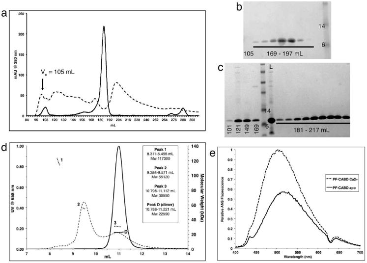
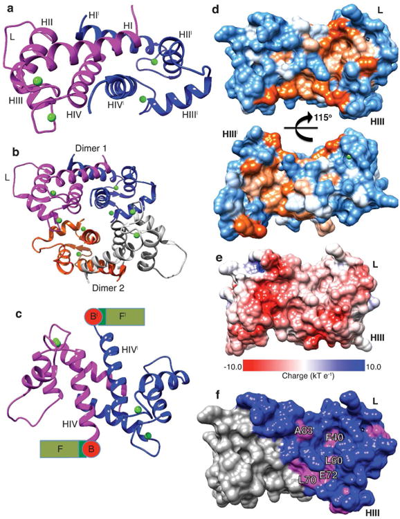
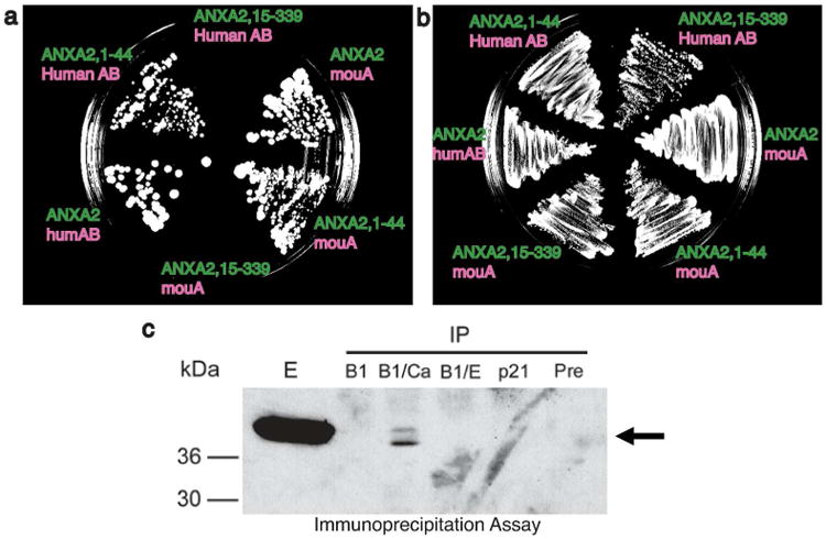
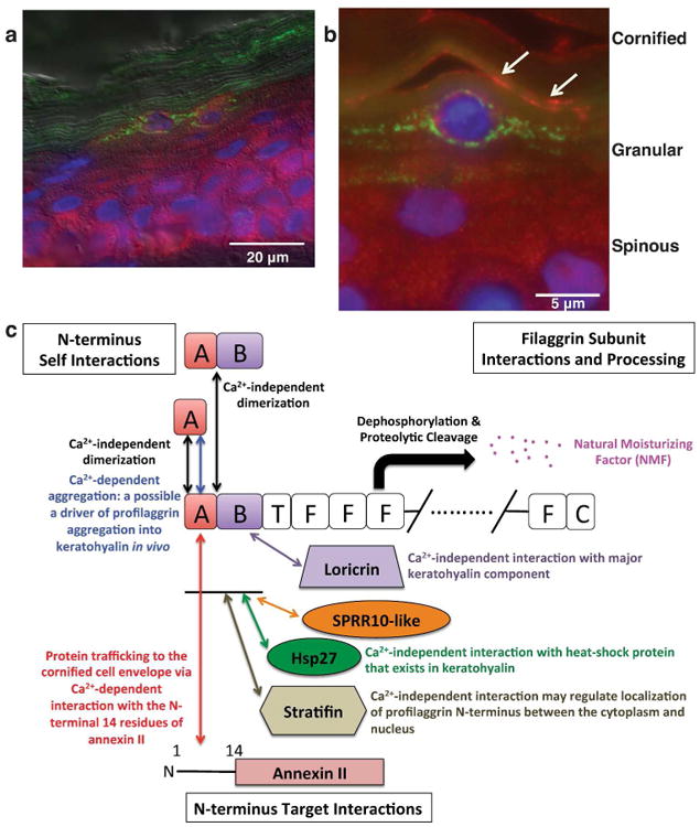
The fused-type S100 protein profilaggrin and its proteolytic products including filaggrin are important in the formation of a normal epidermal barrier; however, the specific function of the S100 calcium-binding domain in profilaggrin biology is poorly understood. To explore its molecular function, we determined a 2.2 Å-resolution crystal structure of the N-terminal fused-type S100 domain of human profilaggrin with bound calcium ions. The profilaggrin S100 domain formed a stable dimer, which contained two hydrophobic pockets that provide a molecular interface for protein interactions. Biochemical and molecular approaches demonstrated that three proteins, annexin II/p36, stratifin/14-3-3 sigma, and heat shock protein 27, bind to the N-terminal domain of human profilaggrin; one protein (stratifin) co-localized with profilaggrin in the differentiating granular cell layer of human skin. Together, these findings suggest a model where the profilaggrin N-terminus uses calcium-dependent and calcium-independent protein-protein interactions to regulate its involvement in keratinocyte terminal differentiation and incorporation into the cornified cell envelope.
Conflict of interest statement
Figures
Similar articles
-
Structural properties of target binding by profilaggrin A and B domains and other S100 fused-type calcium-binding proteins.J Dermatol Sci. 2020 Oct;100(1):39-49. doi: 10.1016/j.jdermsci.2020.08.009. Epub 2020 Aug 21. J Dermatol Sci. 2020. PMID: 32893105 Free PMC article.
-
Functional analysis of the profilaggrin N-terminal peptide: identification of domains that regulate nuclear and cytoplasmic distribution.J Invest Dermatol. 2002 Sep;119(3):661-9. doi: 10.1046/j.1523-1747.2002.01831.x. J Invest Dermatol. 2002. PMID: 12230510
-
Translocation of profilaggrin N-terminal domain into keratinocyte nuclei with fragmented DNA in normal human skin and loricrin keratoderma.Lab Invest. 1998 Oct;78(10):1245-53. Lab Invest. 1998. PMID: 9800950
-
The human epidermal differentiation complex: cornified envelope precursors, S100 proteins and the 'fused genes' family.Exp Dermatol. 2012 Sep;21(9):643-9. doi: 10.1111/j.1600-0625.2012.01472.x. Epub 2012 Apr 16. Exp Dermatol. 2012. PMID: 22507538 Review.
-
Programmed cell death in normal epidermis and loricrin keratoderma. Multiple functions of profilaggrin in keratinization.J Investig Dermatol Symp Proc. 1999 Sep;4(2):145-9. doi: 10.1038/sj.jidsp.5640198. J Investig Dermatol Symp Proc. 1999. PMID: 10536989 Review.
Cited by
-
The X-Ray Crystal Structure of the Keratin 1-Keratin 10 Helix 2B Heterodimer Reveals Molecular Surface Properties and Biochemical Insights into Human Skin Disease.J Invest Dermatol. 2017 Jan;137(1):142-150. doi: 10.1016/j.jid.2016.08.018. Epub 2016 Sep 3. J Invest Dermatol. 2017. PMID: 27595935 Free PMC article.
-
A keratin scaffold regulates epidermal barrier formation, mitochondrial lipid composition, and activity.J Cell Biol. 2015 Dec 7;211(5):1057-75. doi: 10.1083/jcb.201404147. J Cell Biol. 2015. PMID: 26644517 Free PMC article.
-
Cracking the Skin Barrier: Liquid-Liquid Phase Separation Shines under the Skin.JID Innov. 2021 Jul 6;1(3):100036. doi: 10.1016/j.xjidi.2021.100036. eCollection 2021 Sep. JID Innov. 2021. PMID: 34909733 Free PMC article. Review.
-
Skin barrier dysfunction and filaggrin.Arch Pharm Res. 2021 Jan;44(1):36-48. doi: 10.1007/s12272-021-01305-x. Epub 2021 Jan 18. Arch Pharm Res. 2021. PMID: 33462753 Review.
-
Host Responses in an Ex Vivo Human Skin Model Challenged With Malassezia sympodialis.Front Cell Infect Microbiol. 2021 Jan 21;10:561382. doi: 10.3389/fcimb.2020.561382. eCollection 2020. Front Cell Infect Microbiol. 2021. PMID: 33552997 Free PMC article.
References
-
- Aho S, Harding CR, Lee JM, et al. Regulatory role for the profilaggrin N-terminal domain in epidermal homeostasis. J Invest Dermatol. 2012;132:2376–85. - PubMed
-
- Akiyama M. FLG mutations in ichthyosis vulgaris and atopic eczema: spectrum of mutations and population genetics. Br J Dermatol. 2010;162:472–7. - PubMed
Publication types
MeSH terms
Substances
Grants and funding
LinkOut - more resources
Full Text Sources
Other Literature Sources
Research Materials
Miscellaneous
