. 2015 Mar 11;10(3):e0118266.
doi: 10.1371/journal.pone.0118266. eCollection 2015.

A comprehensive library of familial human amyotrophic lateral sclerosis induced pluripotent stem cells

Affiliations

A comprehensive library of familial human amyotrophic lateral sclerosis induced pluripotent stem cells

Ying Li et al. PLoS One. .

Abstract

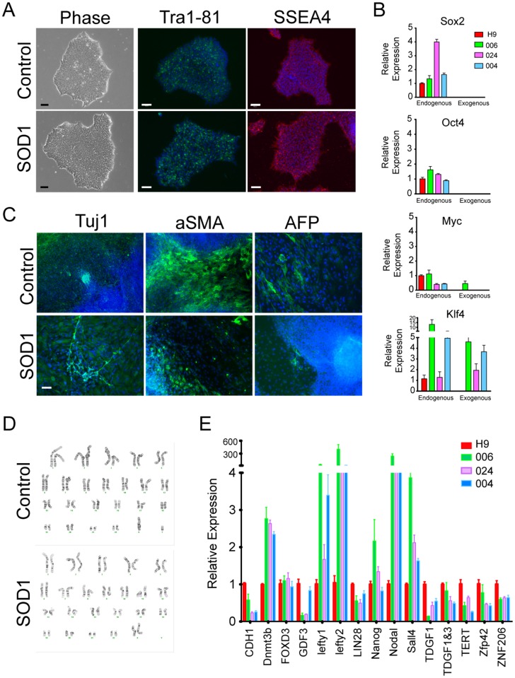
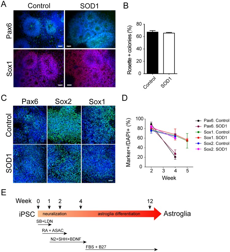
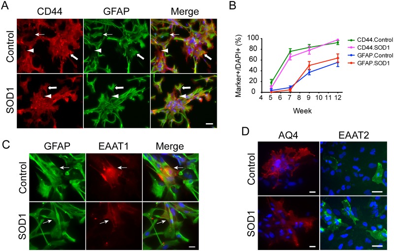
Amyotrophic lateral sclerosis is a progressive disease characterized by the loss of upper and lower motor neurons, leading to paralysis of voluntary muscles. About 10% of all ALS cases are familial (fALS), among which 15-20% are linked to Cu/Zn superoxide dismutase (SOD1) mutations, usually inherited in an autosomal dominant manner. To date only one FDA approved drug is available which increases survival moderately. Our understanding of ALS disease mechanisms is largely derived from rodent model studies, however due to the differences between rodents and humans, it is necessary to have humanized models for studies of disease pathogenesis as well as drug development. Therefore, we generated a comprehensive library of a total 22 of fALS patient-specific induced pluripotent stem cell (iPSC) lines. These cells were thoroughly characterized before being deposited into the library. The library of cells includes a variety of C9orf72 mutations, sod1 mutations, FUS, ANG and FIG4 mutations. Certain mutations are represented with more than one line, which allows for studies of variable genetic backgrounds. In addition, these iPSCs can be successfully differentiated to astroglia, a cell type known to play a critical role in ALS disease progression. This library represents a comprehensive resource that can be used for ALS disease modeling and the development of novel therapeutics.


Conflict of interest statement

Competing Interests: The authors have declared that no competing interests exist.

Figures

Fig 1
Fig 1. Characterization of fALS-iPSCs.
(A) Representative pictures of control- (006) and SOD1-iPSC (002) morphology and their expression of Tra1–81 (green) and SSEA4 (red). Nuclei were stained with DAPI (blue). Size bar: 100μm. (B) Representative qPCR evaluation of endogenous and transgene expression by control- (006) and SOD1-iPSCs (004 and 024). (C) Representative pictures show both control (006) and SOD1-iPSCs (024) generated cells representing the three embryonic germ layers (Tuj1, aSMA and AFP, all in green). Nuclei were stained with DAPI (blue). Size bar: 100μm. (D) Representative karyotypes of control- (006) and SOD1-iPSCs (024). (E) Representative qPCR results of pluripotent marker expression by control (006) and SOD1-iPS (004 and 024) lines.
Fig 2
Fig 2. Differentiation of SOD1-iPSCs to NPCs.
(A, B) NPCs were generated via EB formation. (A) Rosettes and neural tube structures were formed and the cells expressed Pax6 and Sox1. Representative pictures were taken from 006 control and 002 SOD1 lines. Nuclei were stained with DAPI. (B) Quantification of colonies with rosette structures. (C, D) NPCs were induced by inhibition of SMAD pathway. Most cells expressed Pax6, Sox2 and Sox1 at week 2. Nuclei were stained with DAPI. (D) Dynamic examination of Pax6, Sox1 and Sox2 expression by NPCs after 2–5 week. (E) Time line of neural induction by the inhibition of SMAD pathway and astrocyte differentiation. Size bar, 50μm.
Fig 3
Fig 3. Differentiation of SOD1-iPSCs to astroglia.
Representative pictures of control (006) and SOD1-iPSC (008) derived astrocyte after 15-week differentiation showing astroglial marker expression. (A) CD44 (red) and GFAP (green) expression by differentiated cells. Thin arrows indicate GFAP+ only cells. Arrowheads indicate CD44+ only cells. Thick arrows indicate CD44+/GFAP+ cells. (B) Quantification of CD44+ and GFAP+ cells at different time points. (C) EAAT1 (red) expression by GFAP+ (green) astrocytes. Arrows indicate double positive cells. (D) Aquaporin 4 (AQ4, red) and EAAT2 (green) expression. Nuclei were stained with DAPI (blue). Size bar, 20μm.

References

    1. Mulder DW (1982) Clinical limits of amyotrophic lateral sclerosis. Adv Neurol 36: 15–22. - PubMed
    1. Rothstein JD (2009) Current hypotheses for the underlying biology of amyotrophic lateral sclerosis. Ann Neurol 65 Suppl 1: S3–9. 10.1002/ana.21543 - DOI - PubMed
    1. Pasinelli P, Brown RH (2006) Molecular biology of amyotrophic lateral sclerosis: insights from genetics. Nat Rev Neurosci 7: 710–723. - PubMed
    1. DeJesus-Hernandez M, Mackenzie IR, Boeve BF, Boxer AL, Baker M, et al. (2011) Expanded GGGGCC hexanucleotide repeat in noncoding region of C9ORF72 causes chromosome 9p-linked FTD and ALS. Neuron 72: 245–256. 10.1016/j.neuron.2011.09.011 - DOI - PMC - PubMed
    1. Renton AE, Majounie E, Waite A, Simon-Sanchez J, Rollinson S, et al. (2011) A hexanucleotide repeat expansion in C9ORF72 is the cause of chromosome 9p21-linked ALS-FTD. Neuron 72: 257–268. 10.1016/j.neuron.2011.09.010 - DOI - PMC - PubMed

Publication types