. 2015 Mar;71(Pt 3):534-40.
doi: 10.1107/S1399004714027643. Epub 2015 Feb 26.

Automating the application of smart materials for protein crystallization

Affiliations

Automating the application of smart materials for protein crystallization

Sahir Khurshid et al. Acta Crystallogr D Biol Crystallogr. 2015 Mar.

Abstract

The fabrication and validation of the first semi-liquid nonprotein nucleating agent to be administered automatically to crystallization trials is reported. This research builds upon prior demonstration of the suitability of molecularly imprinted polymers (MIPs; known as `smart materials') for inducing protein crystal growth. Modified MIPs of altered texture suitable for high-throughput trials are demonstrated to improve crystal quality and to increase the probability of success when screening for suitable crystallization conditions. The application of these materials is simple, time-efficient and will provide a potent tool for structural biologists embarking on crystallization trials.

Keywords: molecularly imprinted polymers; protein crystallization; smart materials.


Figures

Figure 1
Figure 1
Fabrication of MIPs. Schematic illustration of the steps involved in preparing MIPs for crystallization studies. The initial assembly between the protein template and monomer is advanced through the presence of a polymerizing cross-linker. The protein is then eluted, leaving a protein-specific cavity known as a ‘ghost site’ (adapted from http://www.biotage.com/product-page/mips---molecularly-imprinted-polymers).
Figure 2
Figure 2
Thaumatin screening. Thaumatin crystals grown using a trypsin-MIP. These crystals were obtained using a screen condition that did not yield crystals in control drops [0.2 M ammonium sulfate, 30%(w/v) PEG 8000]. The presence of MIPs facilitated thaumatin crystal formation in a further 14 screen conditions where controls did not form, as detailed in Table 1 ▶. 14 of the 15 conditions did not contain tartrate, which is the most potent precipitant for thaumatin crystallization. Visualization of the modified MIPs is challenging. This is primarily owing to their altered consistency with respect to the original MIPs and also owing to the minute volume being added. Furthermore, if any precipitate forms within the crystallization drops (as in this instance) visualization is more improbable. The scale bar corresponds to 50 µm.
Figure 3
Figure 3
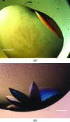
Pgp3 optimization. (a) A large, single Pgp3 crystal grown under metastable conditions using a cognate MIP. Single Pgp3 crystals were obtained using Pgp3-MIPs at dilutions of the original screen ‘hit’ between 50 and 70%. Corresponding controls remained clear. The scale bar corresponds to 75 µm. (b) Clusters of multiple Pgp3 crystals were reproducibly obtained using Pgp3-MIPs when the dilution of the original screen ‘hit’ was greater than 70%. The scale bar corresponds to 100 µm.
Figure 4
Figure 4
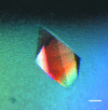
MIF optimization. A human macrophage migration inhibitory factor (MIF) crystal grown using a cognate MIF-MIP. This crystal was grown under metastable conditions (80% dilution of the original ‘hit’ condition) which would not normally yield crystals. The scale bar corresponds to 50 µm.
Figure 5
Figure 5
Nucleation in pores/cavities. Schematic illustration (modified from Frenkel, 2006 ▶) indicating the potential secondary nucleation sites formed when a protein crystal nucleates within a pore or cavity on a generic porous nucleating substrate. According to the theoretical model proposed by Page & Sear (2006 ▶), (a) the initial critical nucleus forms at the corner of the pore, (b) the pore is filled followed by subsequent growth out of the pore and (c) a second critical nucleus forms at the point where the protein aggregate growing out of the pore forms a junction with the nucleant surface. This model is based upon computer simulations, with the white voids observed being a consequence of the simulation process. A critical nucleus can comprise between ten and 100 protein molecules. It is possible that another secondary nucleation site can form at the location indicated by the red arrow. At higher levels of metastability there is sufficient protein to feed both nucleation sites. Furthermore, it is also possible that the protein aggregate growing from the pore may form a crystal itself.

References

    1. Asanithi, P., Saridakis, E., Govada, L., Jurewicz, I., Brunner, E. W., Ponnusamy, R., Cleaver, J. A., Dalton, A. B., Chayen, N. E. & Sear, R. P. (2009). ACS Appl. Mater. Interfaces, 1, 1203–1210. - PubMed
    1. Asherie, N. (2004). Methods, 34, 266–272. - PubMed
    1. Caffrey, M. & Cherezov, V. (2009). Nature Protoc. 4, 706–731. - PMC - PubMed
    1. Chayen, N. E., Saridakis, E. & Sear, R. P. (2006). Proc. Natl Acad. Sci. USA, 103, 597–601. - PMC - PubMed
    1. Curcio, E., Profio, G. D. & Drioli, E. (2003). J. Cryst. Growth, 247, 166–176.

Publication types

MeSH terms