. 2015 Mar;71(Pt 3):565-77.
doi: 10.1107/S1399004714027722. Epub 2015 Feb 26.

Structures of three polycystic kidney disease-like domains from Clostridium histolyticum collagenases ColG and ColH

Affiliations

Structures of three polycystic kidney disease-like domains from Clostridium histolyticum collagenases ColG and ColH

Ryan Bauer et al. Acta Crystallogr D Biol Crystallogr. 2015 Mar.

Abstract

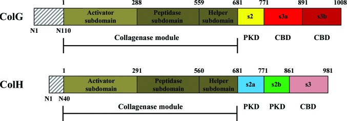
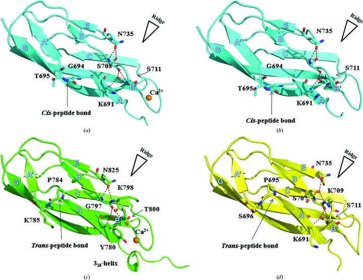
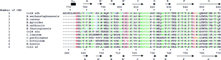
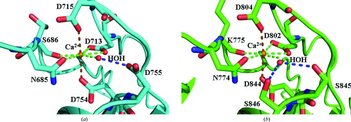
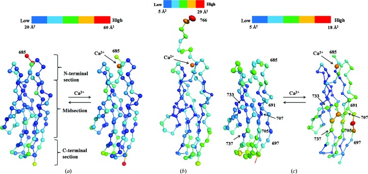
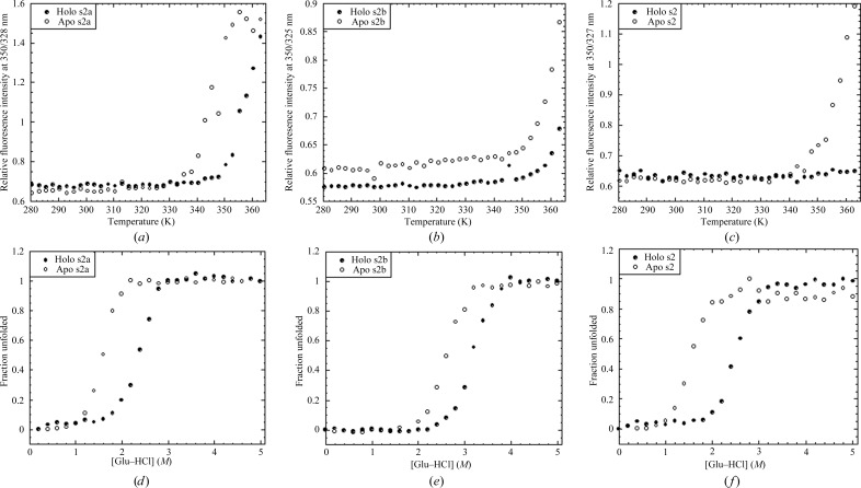
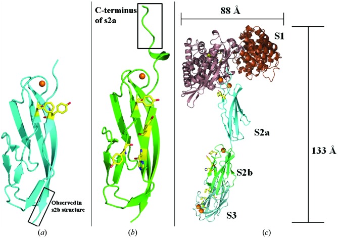
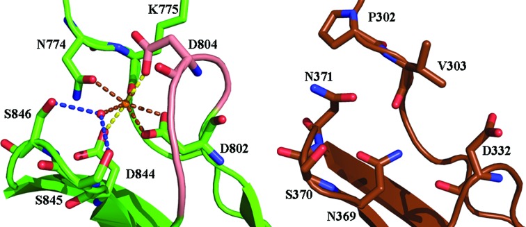
Clostridium histolyticum collagenases ColG and ColH are segmental enzymes that are thought to be activated by Ca(2+)-triggered domain reorientation to cause extensive tissue destruction. The collagenases consist of a collagenase module (s1), a variable number of polycystic kidney disease-like (PKD-like) domains (s2a and s2b in ColH and s2 in ColG) and a variable number of collagen-binding domains (s3 in ColH and s3a and s3b in ColG). The X-ray crystal structures of Ca(2+)-bound holo s2b (1.4 Å resolution, R = 15.0%, Rfree = 19.1%) and holo s2a (1.9 Å resolution, R = 16.3%, Rfree = 20.7%), as well as of Ca(2+)-free apo s2a (1.8 Å resolution, R = 20.7%, Rfree = 27.2%) and two new forms of N-terminally truncated apo s2 (1.4 Å resolution, R = 16.9%, Rfree = 21.2%; 1.6 Å resolution, R = 16.2%, Rfree = 19.2%), are reported. The structurally similar PKD-like domains resemble the V-set Ig fold. In addition to a conserved β-bulge, the PKD-like domains feature a second bulge that also changes the allegiance of the subsequent β-strand. This β-bulge and the genesis of a Ca(2+) pocket in the archaeal PKD-like domain suggest a close kinship between bacterial and archaeal PKD-like domains. Different surface properties and indications of different dynamics suggest unique roles for the PKD-like domains in ColG and in ColH. Surface aromatic residues found on ColH s2a-s2b, but not on ColG s2, may provide the weak interaction in the biphasic collagen-binding mode previously found in s2b-s3. B-factor analyses suggest that in the presence of Ca(2+) the midsection of s2 becomes more flexible but the midsections of s2a and s2b stay rigid. The different surface properties and dynamics of the domains suggest that the PKD-like domains of M9B bacterial collagenase can be grouped into either a ColG subset or a ColH subset. The conserved properties of PKD-like domains in ColG and in ColH include Ca(2+) binding. Conserved residues not only interact with Ca(2+), but also position the Ca(2+)-interacting water molecule. Ca(2+) aligns the N-terminal linker approximately parallel to the major axis of the domain. Ca(2+) binding also increases stability against heat and guanidine hydrochloride, and may improve the longevity in the extracellular matrix. The results of this study will further assist in developing collagen-targeting vehicles for various signal molecules.

Keywords: Clostridium histolyticum; ColG; ColH; polycystic kidney disease-like domains.


Figures

Figure 1
Figure 1
Domain map of collagenases ColG and ColH from C. histolyticum. The pre-pro-peptide (grey hatching) is cleaved from the mature enzyme and indicated by sequence numbering N1–N110 (ColG) and N1–N40 (ColH). The collagenase module is composed of an activator subdomain (olive) and peptidase subdomain (dark olive) that is accompanied by a helper subdomain. The PKD-like domain(s) (yellow for ColG; cyan and green for ColH) connect the collagenase module to the C-­terminal CBD(s) (red for ColG; salmon for ColH) that are responsible for collagen binding.
Figure 2
Figure 2
Structural comparison of holo s2a (a), apo s2a (b), holo s2b (c) and apo s2 (d). Hydrogen bonds that stabilize β-bulges are highlighted. This figure was prepared using PyMOL (Schrödinger).
Figure 3
Figure 3
Structure-based sequence alignment of PKD-like domains from M9B. Residues responsible for Ca2+ binding and for positioning the Ca2+-interacting water, architecturally critical residues and surface aromatic residues are shown in red, orange, green and blue, respectively. Sequence numbering and secondary-structure positions for s2b are shown at the top of the figure. Secondary-structure positions for the s2 structure are similar, although the 310-­helix is absent. Sequence numbering for s2a and s2, as well as secondary-structure positions for s2a, are shown at the bottom of the figure. Sequence alignment was aided by the use of ClustalW2 (Thompson et al., 1994 ▶).
Figure 4
Figure 4
Ca2+ coordination in s2a (a) and s2b (b). Seven O atoms from five residues and one water molecule coordinate to Ca2+ in a pentagonal bipyramidal geometry. Pentagonal base interactions are indicated using brown dashes, while axial interactions are indicated using yellow dashes. Residue-to water interactions are indicated with blue dashes. Either one aspartate (s2a) or adjacent serines (s2b) are responsible for positioning the water molecule along the pentagonal base. This figure was prepared using PyMOL (Schrödinger).
Figure 5
Figure 5
Cα B-factor changes upon Ca2+ binding for s2a (a) and s2 (b). The Cα B factors of holo s2b (c) are also shown. This figure was prepared using PyMOL (Schrödinger).
Figure 6
Figure 6
Results of fluorescence-measured equilibrium denaturation of (a, d) s2a, (b, e) s2b and (c, f) s2 in their apo (open circles) and holo (closed circles) forms.
Figure 7
Figure 7
Surface aromatic residues in s2a (a) and s2b (b). The boxed regions correspond to residues Ala766–Asp770, which are observed in both the s2a and s2b structures and were used to help assemble the full holo ColH structure (c). This structure is assembled from the crystal structures of the peptidase domains of s1, s2a, s2b and s3, as well as the homology-modeled activator domain of s1. Homology modeling was accomplished using SWISS-MODEL (Biasini et al., 2014 ▶). Surface-exposed aromatic residues of the peptidase domain of s1 and s2a-s2b as well as the conserved collagen-interacting aromatic residues of s3 are shown in yellow. Ca2+ is shown as orange spheres. This figure was prepared using PyMOL (Schrödinger).
Figure 8
Figure 8
Proposed evolution of the Ca2+-binding pocket in bacterial PKD-like domains (holo s2b is shown on the left) from the archaeal PKD-like domain (PDB entry 1loq, shown on the right). This figure was prepared using PyMOL (Schrödinger).

References

    1. Adhikari, A. S., Glassey, E. & Dunn, A. R. (2012). J. Am. Chem. Soc. 134, 13259–13265. - PMC - PubMed
    1. Akimoto, M., Takeda, A., Matsushita, O., Inoue, J., Sakamoto, K., Hattori, M., Kounoike, N. & Uchinuma, E. (2013). Plast. Reconstr. Surg. 131, 717–725. - PubMed
    1. Bauer, R., Wilson, J. J., Philominathan, S. T., Davis, D., Matsushita, O. & Sakon, J. (2013). J. Bacteriol. 195, 318–327. - PMC - PubMed
    1. Bertini, I., Fragai, M., Luchinat, C., Melikian, M., Toccafondi, M., Lauer, J. L. & Fields, G. B. (2012). J. Am. Chem. Soc. 134, 2100–2110. - PMC - PubMed
    1. Biasini, M., Bienert, S., Waterhouse, A., Arnold, K., Studer, G., Schmidt, T., Kiefer, F., Cassarino, T. G., Bertoni, M., Bordoli, L. & Schwede, T. (2014). Nucleic Acids Res. 42, W252–W258. - PMC - PubMed

Publication types

MeSH terms

Substances