. 2015 Mar;71(Pt 3):732-41.
doi: 10.1107/S1399004715000978. Epub 2015 Feb 26.

Snapshots of ligand entry, malleable binding and induced helical movement in P-glycoprotein

Affiliations

Snapshots of ligand entry, malleable binding and induced helical movement in P-glycoprotein

Paul Szewczyk et al. Acta Crystallogr D Biol Crystallogr. 2015 Mar.

Abstract

P-glycoprotein (P-gp) is a transporter of great clinical and pharmacological significance. Several structural studies of P-gp and its homologs have provided insights into its transport cycle, but questions remain regarding how P-gp recognizes diverse substrates and how substrate binding is coupled to ATP hydrolysis. Here, four new P-gp co-crystal structures with a series of rationally designed ligands are presented. It is observed that the binding of certain ligands, including an ATP-hydrolysis stimulator, produces a large conformational change in the fourth transmembrane helix, which is positioned to potentially transmit a signal to the nucleotide-binding domains. A new ligand-binding site on the surface of P-gp facing the inner leaflet of the membrane is also described, providing vital insights regarding the entry mechanism of hydrophobic drugs and lipids into P-gp. These results represent significant advances in the understanding of how P-gp and related transporters bind and export a plethora of metabolites, antibiotics and clinically approved and pipeline drugs.

Keywords: P-glycoprotein.


Figures

Figure 1
Figure 1
Structure and function of selenium-labelled homotrimeric cyclopeptides. (a) The chemical structures of the cyclopeptide series bear an identical backbone but with side chains systematically increasing in size and hydrophobicity (from left to right). (b) Stimulation of the basal ATPase activity of purified P-gp. QZ-Ala (green) conferred the highest degree of stimulation (∼16-fold) relative to the basal activity, with an EC50 value of 0.92 µM, followed by QZ-Val (blue; ∼7-fold). Data for verapamil (black), QZ-Leu (yellow) and QZ-Phe (red) are shown. (c) Inhibition of calcein-AM transport in P-gp-overexpressing CR1R12 cells. The same color scheme is applied for each compound as in (b) and the data were fitted using the Hill equation. The mean and SD of triple and quadruplet experiments are shown in (b) and (c), respectively.
Figure 2
Figure 2
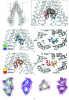
Overview of mouse P-gp at 3.4 Å resolution. Magnified insets at different orientations are shown with the resulting 2mF oDF c electron density (where m is the figure of merit and D is the σA weighting factor) contoured at 1σ; individual transmembrane helices are shown in different colors. TM1 is in red, TM2 is in orange, TM3 is in yellow, TM4 is in light green, TM5 is in sky blue, TM6 is in pink, TM7 is in dark brown, TM8 is in olive, TM9 is in light brown, TM10 is in forest, TM11 is in dark blue and TM12 in deep purple.
Figure 3
Figure 3
Overview of P-gp–cyclopeptide co-crystal structures. (a) Superposition of homotrimeric cyclopeptide compounds bound to P-gp, showing their relative location and orientation in the substrate-binding pocket. (b) Two orientations of subset A ligands (QZ-Ala and QZ-Val; displayed as sticks) bound in the substrate-binding pocket of P-gp. QZ-Ala is shown in green and QZ-Val in blue. (c) Two orientations of subset B ligands (QZ-Leu and QZ-Phe) bound in the substrate-binding pocket of P-gp. QZ-Leu is shown in yellow and QZ-Phe in red. (d) Close-up view of ligands, colored as in (b) and (c), with the resulting 2mF oDF c electron density in blue (contour level of 1.0σ) and anomalous difference density peaks in pink (contour levels of 3.0σ for QZ-Ala, QZ-Va and Q-Phe and 4.0σ for QZ-Leu) for the Se atoms (orange spheres).
Figure 4
Figure 4
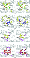
Key P-gp residues involved in binding homotrimeric cyclopeptides. Stereoviews of the binding pocket are shown perpendicular to the membrane and viewed from the cytosol. P-gp residues are shown as sticks, with those involved in binding subset A ligands colored yellow and those involved in binding subset B ligands colored magenta. The ligand coloring is consistent with that shown in Fig. 3 ▶; QZ-Ala, green; QZ-Val, blue; QZ-Leu, yellow; QZ-Phe, red.
Figure 5
Figure 5
Overview of the EH2 ligand-binding site and the ligand-induced movement of TM4. (a) The kinking of TM4 in response to subset A ligands is shown (blue ribbon) in comparison to its ‘straight’ topology in the subset B co-crystal structures (red ribbon). The position of QZ-Val ligands and the resulting 2mF oDF c electron density (contoured at 1.0σ) are displayed. Residues Pro219 and Trp228 are shown as yellow spheres. (b) QZ-Val bound at the EH2 site with P-gp rendered as a molecular-surface representation. The resulting 2mF oDF c electron density for the ligand is shown at a contour level of 1.0σ and the resulting anomalous difference peaks for the Se atoms (orange spheres) are shown at 3.5σ. The close-up inset view depicts the surrounding TM9 and TM12 as brown and purple sticks, respectively, and EH2 as black sticks. Residues in close vicinity to the ligand (Trp694, Phe990 and Tyr994) are labeled.

References

    1. Adams, P. D. et al. (2010). Acta Cryst. D66, 213–221. - PubMed
    1. Afonine, P. V., Grosse-Kunstleve, R. W., Echols, N., Headd, J. J., Moriarty, N. W., Mustyakimov, M., Terwilliger, T. C., Urzhumtsev, A., Zwart, P. H. & Adams, P. D. (2012). Acta Cryst. D68, 352–367. - PMC - PubMed
    1. Aller, S. G., Yu, J., Ward, A., Weng, Y., Chittaboina, S., Zhuo, R., Harrell, P. M., Trinh, Y. T., Zhang, Q., Urbatsch, I. L. & Chang, G. (2009). Science, 323, 1718–1722. - PMC - PubMed
    1. Al-Shawi, M. K. & Omote, H. (2005). J. Bioenerg. Biomembr. 37, 489–496. - PMC - PubMed
    1. Al-Shawi, M. K. & Senior, A. E. (1993). J. Biol. Chem. 268, 4197–4206. - PubMed

Publication types

MeSH terms

Substances