. 2015 May 15;191(10):1126-38.
doi: 10.1164/rccm.201412-2214OC.

Diaphragm muscle fiber weakness and ubiquitin-proteasome activation in critically ill patients

Affiliations

Diaphragm muscle fiber weakness and ubiquitin-proteasome activation in critically ill patients

Pleuni E Hooijman et al. Am J Respir Crit Care Med. .

Abstract

Rationale: The clinical significance of diaphragm weakness in critically ill patients is evident: it prolongs ventilator dependency, and increases morbidity and duration of hospital stay. To date, the nature of diaphragm weakness and its underlying pathophysiologic mechanisms are poorly understood.

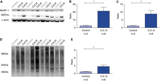
Objectives: We hypothesized that diaphragm muscle fibers of mechanically ventilated critically ill patients display atrophy and contractile weakness, and that the ubiquitin-proteasome pathway is activated in the diaphragm.

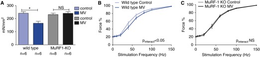
Methods: We obtained diaphragm muscle biopsies from 22 critically ill patients who received mechanical ventilation before surgery and compared these with biopsies obtained from patients during thoracic surgery for resection of a suspected early lung malignancy (control subjects). In a proof-of-concept study in a muscle-specific ring finger protein-1 (MuRF-1) knockout mouse model, we evaluated the role of the ubiquitin-proteasome pathway in the development of contractile weakness during mechanical ventilation.

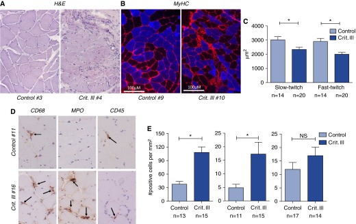
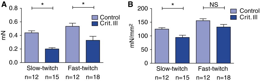
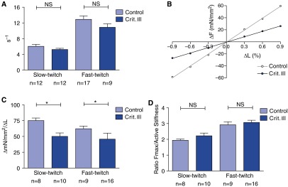
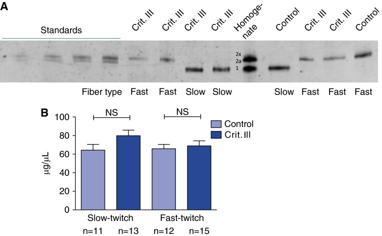
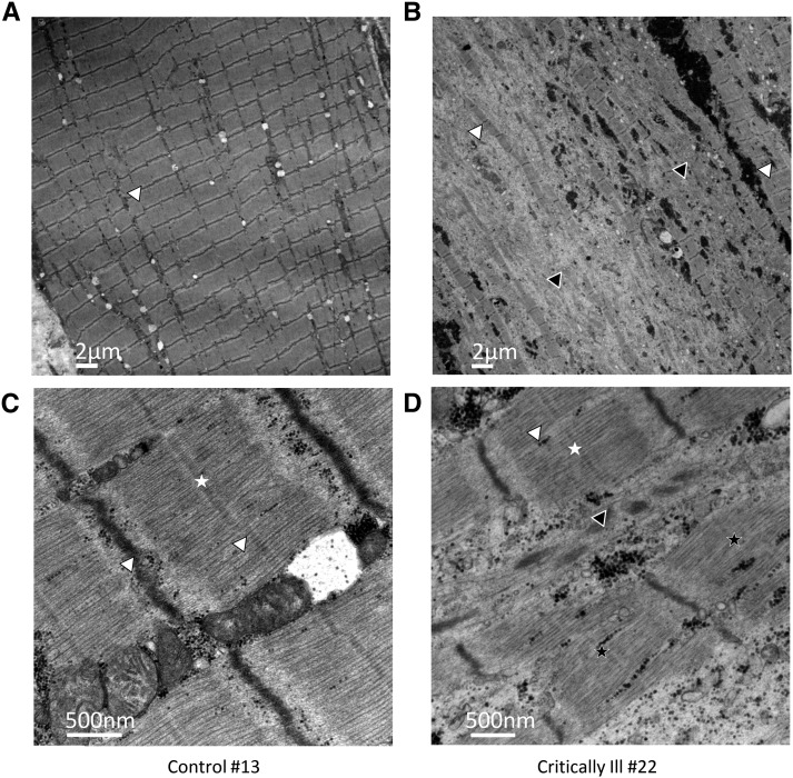
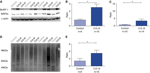
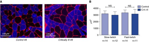
Measurements and main results: Both slow- and fast-twitch diaphragm muscle fibers of critically ill patients had approximately 25% smaller cross-sectional area, and had contractile force reduced by half or more. Markers of the ubiquitin-proteasome pathway were significantly up-regulated in the diaphragm of critically ill patients. Finally, MuRF-1 knockout mice were protected against the development of diaphragm contractile weakness during mechanical ventilation.

Conclusions: These findings show that diaphragm muscle fibers of critically ill patients display atrophy and severe contractile weakness, and in the diaphragm of critically ill patients the ubiquitin-proteasome pathway is activated. This study provides rationale for the development of treatment strategies that target the contractility of diaphragm fibers to facilitate weaning.

Keywords: diaphragm weakness; mechanical ventilation; single muscle fiber; weaning failure.


Figures

Figure 1.
Figure 1.
Atrophy of diaphragm fibers in critically ill patients. (A) Representative examples of hematoxylin and eosin (H&E) staining. (B) Immunohistochemically stained cross-sections of diaphragm muscle fiber biopsies. Red: wheat germ agglutinin indicating membranes. Blue: MY32 antibody specific for fast-twitch fibers. (C) Diaphragm fiber cross-sectional area (μm2) was significantly smaller in slow- and fast-twitch fibers of critically ill patients, indicating atrophy. (D) Representative examples of staining for macrophages (CD68), neutrophil granulocytes (myeloperoxidase [MPO]), and lymphocytes (CD45). (E) In critically ill patients, the number of macrophages and neutrophil granulocytes was significantly higher compared with control subjects. MyHC = myosin heavy chain; NS = not significant. *P < 0.05. Arrows indicate inflammatory cells.
Figure 2.
Figure 2.
Contractile weakness of individual diaphragm fibers in critically ill patients. (A) The maximal absolute force generation of both slow- and fast-twitch diaphragm muscle fibers of critically ill patients was significantly lower compared with control patients. (B) Maximal tension, which is the absolute maximal force normalized to fiber cross-sectional area, was significantly lower in slow-twitch fibers of critically ill patients compared with control fibers, and fast-twitch fibers showed a trend toward a lower maximal tension. NS = not significant. *P < 0.05.
Figure 3.
Figure 3.
Cross-bridge cycling kinetics. (A) The rate constant of force redevelopment did not differ between control and critically ill patients in both fiber types, which suggests there is no reduction in the fraction of strongly bound cross-bridges. (B) Amplitude of the force response (normalized to cross-sectional area) during fiber shortening and lengthening; the slope of this line is the normalized active stiffness (shown are representative single diaphragm fibers of critically ill patient 1 and control patient 1). (C) Normalized active stiffness was significantly lower in slow- and fast-twitch fibers of critically ill patients. (D) The ratio of maximal tension to normalized active stiffness, which is a measure of the force generated per cross-bridge, did not differ between critically ill and control patients. NS = not significant. *P < 0.05.
Figure 4.
Figure 4.
Myosin heavy chain (MyHC) concentration is preserved in diaphragm fibers of critically ill patients. (A) Example of sodium dodecyl sulfate–gel electrophoresis with known amounts of rabbit MyHC (M-1636 Sigma) to determine the amount of MyHC per fiber volume. (B) MyHC concentration of slow- and fast-twitch fibers did not differ between control and critically ill patients. NS = not significant.
Figure 5.
Figure 5.
Ultrastructural damage in diaphragm fibers of critically ill patients. Electron microscopy images of longitudinal sections of diaphragm muscle fibers. (A and C) Control patient; (B and D) critically ill patient. The diaphragm samples of the control patient show intact structure with mostly well-aligned myofibrils. The diaphragm sample of the critically ill patient shows myofibrils that are disarranged (white arrowheads and black arrowheads indicate, respectively, well-arranged and disarranged myofibrils and z-disks; white stars and black stars indicate, respectively, well-arranged and disarranged M-lines).
Figure 6.
Figure 6.
Increased activation of the ubiquitin–proteasome pathway in diaphragm fibers of critically ill patients. (A) Example of Western blot; each lane represents diaphragm muscle homogenates of control and critically ill patients with antibodies for the ubiquitin ligases muscle-specific ring finger protein-1 (MuRF-1) (top), muscle atrophy F-box (MAFbx) (middle), and α-actin (bottom, loading control). In critically ill patients compared with control subjects, there was a significant increase in protein levels of MuRF-1 (twofold, B) and MAFbx (more than threefold, C) relative to α-actin (note that each sample was normalized to a reference control sample that was run on each gel). (D) Example of a Western blot with an antiubiquitin antibody to detect ubiquitinated proteins; each lane represents a diaphragm muscle homogenate of a control or a critically ill patient. (E) The relative level of ubiquitinated proteins was more than fourfold higher in critically ill patients compared with control subjects. *P < 0.05.
Figure 7.
Figure 7.
Intact diaphragm muscle strength in muscle-specific ring finger protein-1 (MuRF-1) knockout (KO) mice. Intact diaphragm muscle strips were isolated from wild-type and MuRF-1 KO mice. (A) Maximal tetanic force was significantly lower in wild-type mice that received mechanical ventilation (MV) compared with wild-type mice that did not receive MV (control subjects). Diaphragm muscle strength of MV MuRF-1 KO mice did not differ from control MuRF-1 KO mice. (B) Compared with the control group, in MV wild-type mice a rightward shift of the force–stimulation frequency relation was present; (C) this rightward shift was not present in MV MuRF-1 KO mice. NS = not significant. *P < 0.05.
Figure 8.
Figure 8.
Noninspiratory muscles of critically ill patients do not exhibit fiber atrophy. (A) Representative examples of immunohistochemically stained cross-sections of biopsies of latissimus dorsi muscle. Red: wheat germ agglutinin indicating membranes. Blue: MY32 antibody specific for fast-twitch fibers. (B) Fiber cross-sectional area (μm2) of noninspiratory muscles did not differ between control and critically ill patients. NS = not significant.
Figure 9.
Figure 9.
Increased activation of the ubiquitin–proteasome pathway in noninspiratory muscles of critically ill patients. (A) Example of a Western blot. Each lane represents diaphragm muscle homogenates of control and critically ill patients with antibodies for the ubiquitin ligases muscle-specific ring finger protein-1 (MuRF-1) (top), muscle atrophy F-box (MAFbx) (middle), and α-actin (bottom, loading control). In critically ill patients compared with control subjects, there was a significant increase in protein levels of MuRF-1 (more than sevenfold, B) and MAFbx (more than ninefold, C) relative to α-actin (note that each sample was normalized to a reference control sample that was run on each gel). (D) Example of a Western blot with an antiubiquitin antibody to detect ubiquitinated proteins; each lane represents a diaphragm muscle homogenate of a control or a critically ill patient. (E) The relative level of ubiquitinated proteins was more than fourfold higher in critically ill patients compared with control subjects. *P < 0.05.

Comment in

  • Muscle weakness in critical illness.
    Sieck GC. Sieck GC. Am J Respir Crit Care Med. 2015 May 15;191(10):1094-6. doi: 10.1164/rccm.201503-0478ED. Am J Respir Crit Care Med. 2015. PMID: 25978566 No abstract available.

References

    1. Batt J, Dos Santos CC, Herridge MS. Muscle injury during critical illness. JAMA. 2013;310:1569–1570. - PubMed
    1. Schweickert WD, Hall J. ICU-acquired weakness. Chest. 2007;131:1541–1549. - PubMed
    1. Herridge MS, Tansey CM, Matté A, Tomlinson G, Diaz-Granados N, Cooper A, Guest CB, Mazer CD, Mehta S, Stewart TE, et al. Canadian Critical Care Trials Group. Functional disability 5 years after acute respiratory distress syndrome. N Engl J Med. 2011;364:1293–1304. - PubMed
    1. De Jonghe B, Bastuji-Garin S, Sharshar T, Outin H, Brochard L. Does ICU-acquired paresis lengthen weaning from mechanical ventilation? Intensive Care Med. 2004;30:1117–1121. - PubMed
    1. Boles J-M, Bion J, Connors A, Herridge M, Marsh B, Melot C, Pearl R, Silverman H, Stanchina M, Vieillard-Baron A, et al. Weaning from mechanical ventilation. Eur Respir J. 2007;29:1033–1056. - PubMed

Publication types

MeSH terms