. 2015;7(2):311-21.
doi: 10.1080/19420862.2015.1007813.

Antibody-mediated phagocytosis contributes to the anti-tumor activity of the therapeutic antibody daratumumab in lymphoma and multiple myeloma

Affiliations

Antibody-mediated phagocytosis contributes to the anti-tumor activity of the therapeutic antibody daratumumab in lymphoma and multiple myeloma

Marije B Overdijk et al. MAbs. 2015.

Abstract

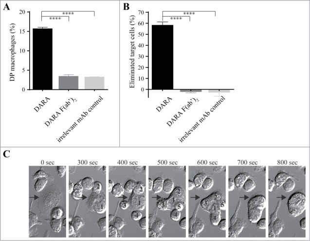
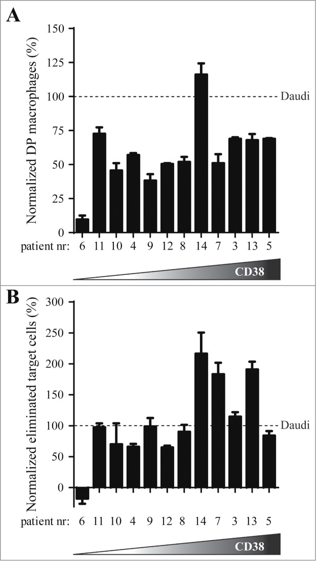
Daratumumab (DARA) is a human CD38-specific IgG1 antibody that is in clinical development for the treatment of multiple myeloma (MM). The potential for IgG1 antibodies to induce macrophage-mediated phagocytosis, in combination with the known presence of macrophages in the tumor microenvironment in MM and other hematological tumors, led us to investigate the contribution of antibody-dependent, macrophage-mediated phagocytosis to DARA's mechanism of action. Live cell imaging revealed that DARA efficiently induced macrophage-mediated phagocytosis, in which individual macrophages rapidly and sequentially engulfed multiple tumor cells. DARA-dependent phagocytosis by mouse and human macrophages was also observed in an in vitro flow cytometry assay, using a range of MM and Burkitt's lymphoma cell lines. Phagocytosis contributed to DARA's anti-tumor activity in vivo, in both a subcutaneous and an intravenous leukemic xenograft mouse model. Finally, DARA was shown to induce macrophage-mediated phagocytosis of MM cells isolated from 11 of 12 MM patients that showed variable levels of CD38 expression. In summary, we demonstrate that phagocytosis is a fast, potent and clinically relevant mechanism of action that may contribute to the therapeutic activity of DARA in multiple myeloma and potentially other hematological tumors.

Keywords: ADCC, antibody-dependent cellular cytotoxicity; BL, Burkitt's lymphoma; BM, bone marrow; Burkitt's lymphoma; CCS, cosmic calf serum; CD38; CDC, complement-dependent cytotoxicity; DARA, daratumumab; DP, double positive; E:T, effector to target ratio; FcγR, Fc-gamma receptor; IMiD, immunomodulatory drug; MM, multiple myeloma; MNC, mononuclear cells; Mϕ, macrophage; PBMC, peripheral blood mononuclear cells; daratumumab; mAb, monoclonal antibody; macrophage; multiple myeloma; phagocytosis; therapeutic antibody.


Figures

Figure 1.
Figure 1.
Flow cytometry and live cell imaging reveals macrophage-mediated phagocytosis of CD38+ tumor cells in the presence of DARA. Co-cultures of mouse mφ and Daudi cells in the presence of 6.7 nM DARA or F(ab’)2 fragments thereof, E:T ratio of 1:1 (A, B) or 3:1 (C). (A) Double positive (DP) mφ were characterized as F4/80+calcein+CD19 and the percentage DP macrophages was calculated as described in Materials and Methods. (B) The percentage eliminated target cells was calculated from the number of remaining F4/80- cells as described in Materials & Methods. Each bar shows mean ± SEM, results from a representative experiment are shown (n = 3). (C) Time-lapse imaging microscopy, bright field images of a mouse mφ (arrow) that sequentially engulfed 5 individual Daudi cells (numbers) over a period of 800 s. The images are representative for observations in multiple independent phagocytosis experiments (n = 3) (****p < 0.0001 Bonferroni's multiple comparison test).
Figure 2.
Figure 2.
Induction of macrophage-mediated phagocytosis is strongly impaired for an IgG2 isotype variant of DARA. Phagocytosis of Daudi cells by mouse mφ in the presence of 6.7 nM mAb, E:T ratio of 1:1. (A) Double-positive (DP) mφ were characterized as F4/80+calcein+CD19 and the percentage DP macrophages was calculated as described in Materials and Methods. (B) Percentage eliminated target cells was calculated using the number of remaining F4/80- cells as described in Materials & Methods. Each bar shows mean ± SEM, results from a representative experiment (n = 3) (**p < 0.01, ****p < 0.0001 Bonferroni's multiple comparison test).
Figure 3.
Figure 3.
Phagocytosis contributes to the in vivo anti-tumor effect of DARA. (A) Kaplan-Meier plot showing time to tumor progression (cutoff set at a tumor volume > 800 mm3) for mice that had been inoculated s.c. with 20 × 106 Daudi-luc cells (8 mice per group). Subsequently, mice were treated i.p. with 250 μg mAb per mouse (∼12.5 mg/kg) at day 0. Tumor progression was significantly reduced in DARA-K322A-treated mice compared to DARA-IgG2-K322A treatment (p < 0.004 Mantle-Cox log-rank test at time to progression). (B) Kaplan-Meier plot showing time to tumor progression (cutoff set at bioluminescence > 50 000 cpm) for mice that had been inoculated i.v. with 2.5×10⁶ Daudi-luc cells (10 mice per group). Subsequently, mice were treated i.p. with 10 μg mAb per mouse (∼0.5 mg/kg) at day 0. Tumor progression was significantly reduced in DARA-K322A-treated vs. DARA-IgG2-K322A-treated mice (p < 0.001 Mantle-Cox log-rank test at time to progression).
Figure 4.
Figure 4.
Human macrophages induce DARA-dependent phagocytosis of BL and MM cell lines. Phagocytosis of BL cells (left panel) or MM cells (right panel) by human mφ in the presence of 6.7 nM mAb, E:T ratio of 2:1. (A) Percentage of double positive (DP) mφ characterized as CD11b+calcein+target Ag. (B) Percentage of eliminated target cells calculated from the number of remaining CD11b- cells as described in Materials & Methods. Each bar shows mean ± SEM, results from a representative experiment (n = 3) (**p < 0.01, ***p < 0.001, ****p < 0.0001 Bonferroni's multiple comparison test).
Figure 5.
Figure 5.
DARA-dependent phagocytosis of patient MM cells by human macrophages. Patient MM cells were incubated with human mφ, obtained from healthy donors (E:T ratio 2:1), in the presence of 6.7 nM mAb. To correct for differences in potency between batches of human donor mφ obtained from different donors, results were normalized by calculating the ratio of patient MM cells to an internal Daudi cell standard (observed in the same experiment). The 12 patient samples are ranked according to their CD38 expression level, with cells from patient 6 exhibiting lowest (10,000 molecules per cells) and patient 5 highest (550,000 molecules/cell) CD38 expression. (A) Normalized percentage of DARA-specific double positive (DP) mφ. (B) Normalized percentage of DARA-specific eliminated target cells. Each bar shows mean ± SEM of triplicates.

References

    1. Manches O, Lui G, Chaperot L, Gressin R, Molens JP, Jacob MC, Sotto JJ, Leroux D, Bensa JC, Plumas J. In vitro mechanisms of action of rituximab on primary non-Hodgkin lymphomas. Blood 2003; 101:949-54; PMID:12393572; http://dx.doi.org/10.1182/blood-2002-02-0469 - DOI - PubMed
    1. Munn DH, McBride M, Cheung NK. Role of low-affinity Fc receptors in antibody-dependent tumor cell phagocytosis by human monocyte-derived macrophages. Cancer Res 1991; 51:1117-23; PMID:1825476 - PubMed
    1. Mantovani A, Schioppa T, Porta C, Allavena P, Sica A. Role of tumor-associated macrophages in tumor progression and invasion. Cancer Metastasis Rev 2006; 25:315-22; PMID:16967326; http://dx.doi.org/10.1007/s10555-006-9001-7 - DOI - PubMed
    1. Ribatti D, Moschetta M, Vacca A. Macrophages in multiple myeloma. Immunol Lett 2014; 161:241–4; PMID:24370642. - PubMed
    1. Zheng Y, Cai Z, Wang S, Zhang X, Qian J, Hong S, Li H, Wang M, Yang J, Yi Q. Macrophages are an abundant component of myeloma microenvironment and protect myeloma cells from chemotherapy drug-induced apoptosis. Blood 2009; 114:3625-8; PMID:19710503; http://dx.doi.org/10.1182/blood-2009-05-220285 - DOI - PMC - PubMed

Publication types