. 2015 May;35(5):1237-45.
doi: 10.3892/ijmm.2015.2128. Epub 2015 Mar 6.

The inhibitory effects of Geranium thunbergii on interferon-γ- and LPS-induced inflammatory responses are mediated by Nrf2 activation

Affiliations

The inhibitory effects of Geranium thunbergii on interferon-γ- and LPS-induced inflammatory responses are mediated by Nrf2 activation

Hee-Jin Choi et al. Int J Mol Med. 2015 May.

Abstract

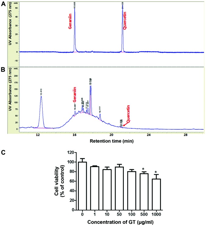
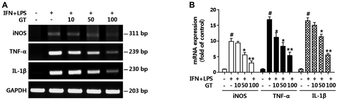
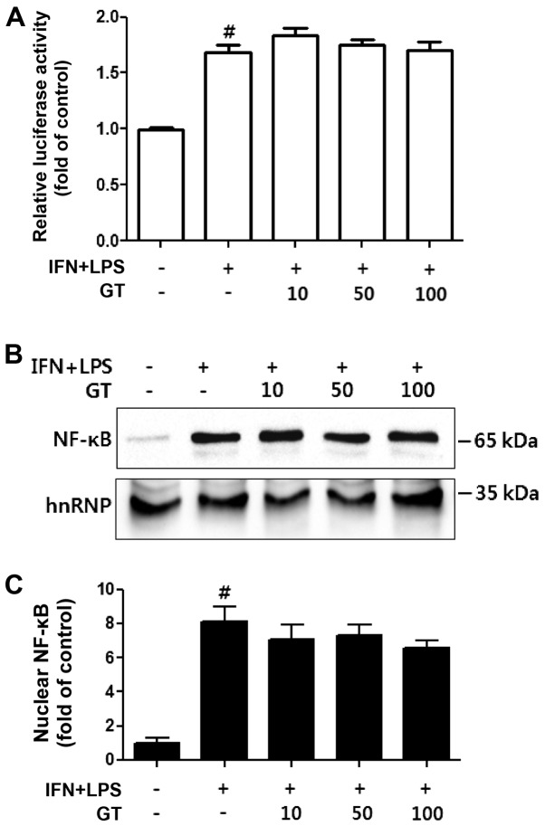
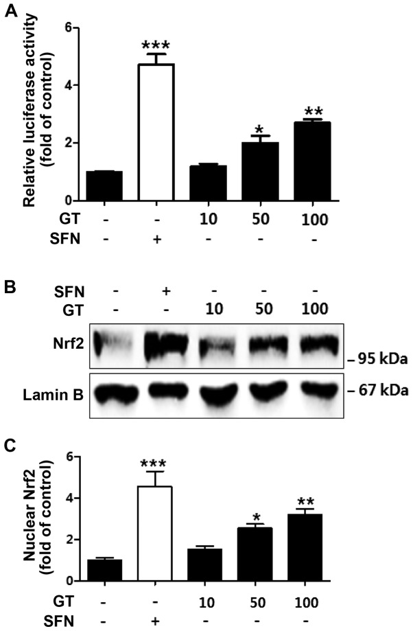
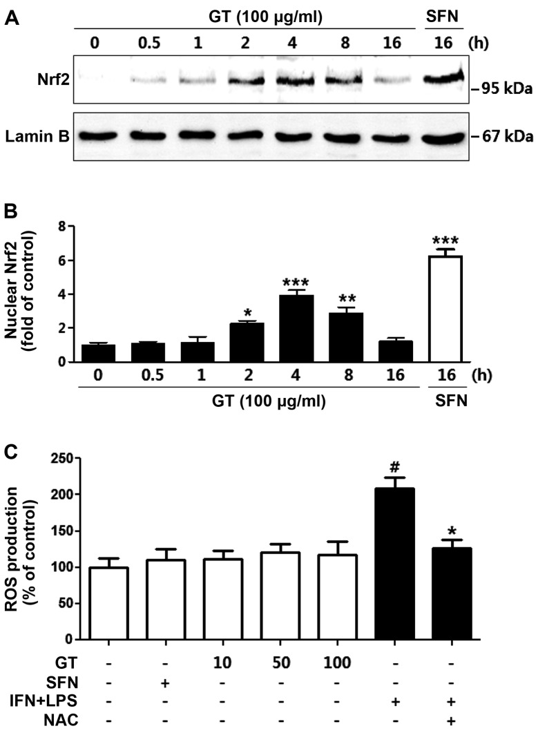
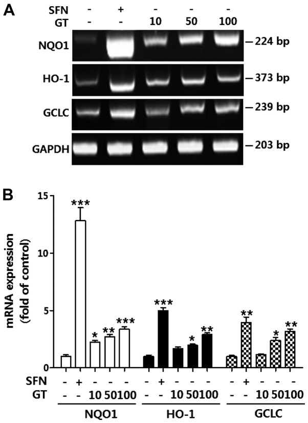
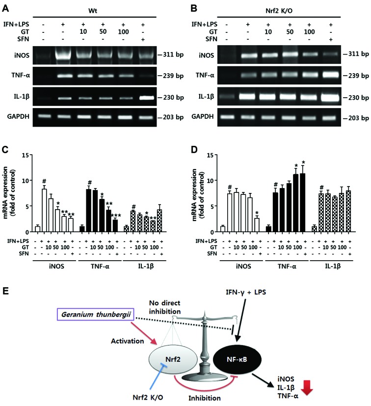
Geranium thunbergii Sieb. et Zucc. (GT; which belongs to the Geraniaceae family) has been used as a traditional medicine in East Asia for the treatment of inflammatory diseases, including arthritis and diarrhea. However, the underlying mechanisms of the anti-inflammatory effects of GT remain poorly understood. In the present study, we examined the mechanisms responsible for the anti-inflammatory activity of GT in macrophages. The results revealed that GT significantly inhibited the lipopolysaccharide (LPS)- and interferon-γ (IFN-γ)-induced expression of pro-inflammatory genes, such as inducible nitric oxide synthase, tumor necrosis factor-α and interleukin-1β, as shown by RT-PCR. However, the inhibitory effects of GT on LPS- and IFN-γ-induced inflammation were associated with an enhanced nuclear factor erythroid 2-related factor 2 (Nrf2) activity, but not with the suppression of nuclear factor (NF)-κB activity, as shown by western blot analysis. In addition, in bone marrow-derived macrophages (BMDM) isolated from Nrf2 knockout mice, GT did not exert any inhibitory effect on the LPS- and IFN-γ-induced inflammation. Taken together, our findings indicate that the anti-inflammatory effects of GT may be associated with the activation of Nrf2, an anti-inflammatory transcription factor.


Figures

Figure 1
Figure 1
Chromatographic analysis and cytotoxic effects of Geranium thunbergii (GT). (A) HPLC chromatogram of mixed standard solutions, geraniin and quercetin. (B) HPLC chromatogram of 50% aquaeous-ethanol extract of GT. (C) RAW264.7 cells were treated with the indicated concentrations of GT for 24 h, and the viability of the cells was examined by MTT assay. The data from 3 independent experiments are represented as the means ± SD. *p<0.05 compared to the controls.
Figure 2
Figure 2
Geranium thunbergii (GT) suppresses the expression of pro-inflammatory genes. RAW264.7 cells were treated with the indicated concentrations of GT for 4 h, prior to stimulation with interferon-γ (IFN-γ) and lipopolysaccharides (LPS) (10 ng/ml, respectively). The cells were cultured for 16 h. (A) The expression of pro-inflammatory genes, includinginducible nitric oxide synthase (iNOS), tumor necrosis factor-α (TNF-α), and interleukin-1β (IL-1β) was evaluated by RT-PCR. The expression of glyceraldehyde-3-phosphate dehydrogenase (GAPDH) was used as an internal control. (B) The intensity of the bands was densitometrically quantified and calculated as the mean ± SD of 3 independent experiments. #p<0.01 compared to the negative control (first column); *p<0.05 and **p<0.01 compared to the positive control (second column).
Figure 3
Figure 3
Geranium thunbergii (GT) does not directly inhibit the transcriptional activity and nuclear translocation of nuclear factor-κB (NF-κB) in RAW264.7 cells. (A) An NF-κB reporter cell line, derived from RAW264.7 cells, was pre-treated with the indicated concentrations of GT for 30 min, then stimulated with interferon-γ (IFN-γ) and lipopolysaccharides (LPS) (10 ng/ml, respectively) for 16 h. #p<0.01 compared to the negative control (first column). (B) The amount of nuclear p65 subunit of NF-κB was examined by western blot analysis. The amount of hnRNP was used as an internal control. (C) The intensity of the bands was densitometrically quantified and calculated as the mean ± SD of 3 independent experiments. #p<0.01 compared to the negative control (first column).
Figure 4
Figure 4
Geranium thunbergii (GT) enhances the transcriptional activity and nuclear translocation of nuclear factor erythroid 2-related factor 2 (Nrf2) in RAW264.7 cells. (A) An Nrf2 reporter cell line was treated with sulforaphane (SFN; 5 μM) or the indicated amounts of GT for 16 h. Luciferase activity was normalized by the amount of total proteins. *p<0.05, **p<0.01 and ***p<0.001 compared to the control (first column). All data represent the means ± SD of 3 independent experiments. (B) The cells were treated with the indicated concentrations of GT for 8 h. SFN (5 μM) was used as a positive control. The amount of nuclear Nrf2 was estimated by western blot analysis. The amount of lamin B was used as an internal control. (C) The intensity of the bands was densitometrically quantified and calculated as the means ± SD of 3 independent experiments. *p<0.05, **p<0.01 and ***p<0.001 compared to the control (first column).
Figure 5
Figure 5
Effect of Geranium thunbergii (GT) on the time-dependent nuclear translocation of nuclear factor erythroid 2-related factor 2 (Nrf2) and production of intracellular reactive oxygen species (ROS). (A) The RAW264.7 cells were treated with 100 μg/ml of GT for the indicated periods of time. The amount of nuclear Nrf2 was examined by western blot analysis. The amount of lamin B was used as an internal control. (B) The intensity of the bands was densitometrically quantified and calculated as the means ± SEM of 3 independent experiments. *p<0.05, **p<0.01 and ***p<0.001 compared to the control (first column). (C) The RAW264.7 cells were treated with the indicated concentrations of GT or sulforaphane (SFN; 5 μM) for 16 h. The cells treated with interferon-γ (IFN-γ) and lipopolysaccharides (LPS) (10 ng/ml, respectively) and/or N-acetyl cysteine (NAC) (1 mM; used as a control). The production of intracellular ROS was estimated using carboxy-H2DCFDA. The data from 3 independent experiments are represented as the means ± SD. #p<0.01 compared to the negative control (first column); *p<0.05 compared to the positive control (sixth column).
Figure 6
Figure 6
Geranium thunbergii (GT) enhances the expression of nuclear factor erythroid 2-related factor 2 (Nrf2)-dependent genes. (A) The RAW264.7 cells were treated with the indicated concentrations of GT for 16 h. sulforaphane (SFN; 5 μM) was used as a positive control. The expression of Nrf2-dependent genes, including NAD(P)H: quinine oxidoreductase 1 (NQO1), heme oxygenase-1 (HO-1) and glutamate-cysteine ligase catalytic subunit (GCLC), were evaluated by RT-PCR. The expression of glyceraldehyde-3-phosphate dehydrogenase (GAPDH) was used as an internal control. (B) The intensity of the bands was densitometrically quantified and calculated as the means ± SD of 3 independent experiments. *p<0.05, **p<0.01 and ***p<0.001 compared to the control (first column).
Figure 7
Figure 7
Inhibitory effects of Geranium thunbergii (GT) on the expression of pro-inflammatory genes are regulated by nuclear factor erythroid 2-related factor 2 (Nrf2) activity. (A and B) Bone marrow-derived macrophages from (A) wild-type C57BL/6 or (B) Nrf2 knockout mice were treated with GT (100 μg/ml) or sulforaphane (SFN; 5 μM) for 4 h prior to stimulation with interferon-γ (IFN-γ) and lipopolysaccharides (LPS) (10 ng/ml, respectively). The expression of pro-inflammatory genes, including inducible nitric oxide synthase (iNOS), interleukin-1β (IL-1β) and tumor necrosis factor-α (TNF-α) were estimated by RT-PCR. The expression of glyceraldehyde-3-phosphate dehydrogenase (GAPDH) was used as an internal control. (C and D) The intensity of the bands was densitometrically quantitated and calculated as the mean ± SD of 3 independent experiments. #p<0.001 compared to the negative control (first column); *p<0.05, **p<0.01 and ***p<0.001 compared to the positive control (second column). (E) A schematic representation of the anti-inflammatory effects of GT.

References

    1. Reuter S, Gupta SC, Chaturvedi MM, Aggarwal BB. Oxidative stress, inflammation, and cancer: How are they linked? Free Radic Biol Med. 2010;49:1603–1616. doi: 10.1016/j.freeradbiomed.2010.09.006. - DOI - PMC - PubMed
    1. Nathan C, Ding A. Nonresolving inflammation. Cell. 2010;140:871–882. doi: 10.1016/j.cell.2010.02.029. - DOI - PubMed
    1. Laskin DL, Sunil VR, Gardner CR, Laskin JD. Macrophages and tissue injury: Agents of defense or destruction? Annu Rev Pharmacol Toxicol. 2011;51:267–288. doi: 10.1146/annurev.pharmtox.010909.105812. - DOI - PMC - PubMed
    1. Davignon JL, Hayder M, Baron M, Boyer JF, Constantin A, Apparailly F, Poupot R, Cantagrel A. Targeting monocytes/macrophages in the treatment of rheumatoid arthritis. Rheumatology (Oxford) 2013;52:590–598. doi: 10.1093/rheumatology/kes304. - DOI - PubMed
    1. Alam J, Stewart D, Touchard C, Boinapally S, Choi AM, Cook JL. Nrf2, a Cap‘n’Collar transcription factor, regulates induction of the heme oxygenase-1 gene. J Biol Chem. 1999;274:26071–26078. doi: 10.1074/jbc.274.37.26071. - DOI - PubMed

Publication types

MeSH terms