. 2015 Jun;24(6):987-95.
doi: 10.1002/pro.2678. Epub 2015 Apr 2.

Cryo-EM structure of fatty acid synthase (FAS) from Rhodosporidium toruloides provides insights into the evolutionary development of fungal FAS

Affiliations

Cryo-EM structure of fatty acid synthase (FAS) from Rhodosporidium toruloides provides insights into the evolutionary development of fungal FAS

Manuel Fischer et al. Protein Sci. 2015 Jun.

Abstract

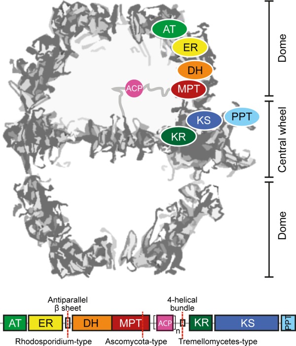
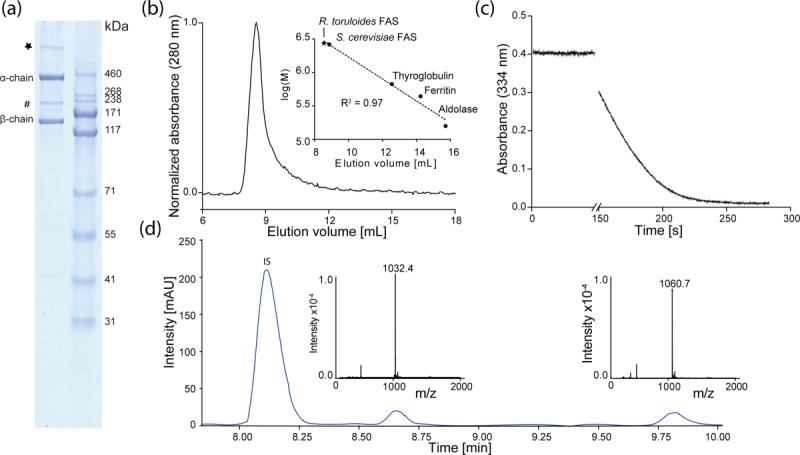
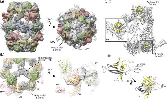
Fungal fatty acid synthases Type I (FAS I) are up to 2.7 MDa large molecular machines composed of large multifunctional polypeptides. Half of the amino acids in fungal FAS I are involved in structural elements that are responsible for scaffolding the elaborate barrel-shaped architecture and turning fungal FAS I into highly efficient de novo producers of fatty acids. Rhodosporidium toruloides is an oleaginous fungal species and renowned for its robust conversion of carbohydrates into lipids to over 70% of its dry cell weight. Here, we use cryo-EM to determine a 7.8-Å reconstruction of its FAS I that reveals unexpected features; its novel form of splitting the multifunctional polypeptide chain into the two subunits α and β, and its duplicated ACP domains. We show that the specific distribution into α and β occurs by splitting at one of many possible sites that can be accepted by fungal FAS I. While, therefore, the specific distribution in α and β chains in R. toruloides FAS I is not correlated to increased protein activities, we also show that the duplication of ACP is an evolutionary late event and argue that duplication is beneficial for the lipid overproduction phenotype.

Keywords: acyl carrier protein; biofuel; mega-enzyme; multifunctional proteins; protein assembly.


Figures

Figure 1
Figure 1
Architecture and domain organization of fungal FAS I. Fungal FAS I is an overall D3 symmetric barrel-shaped complex of homohexameric or heterodocameric (α6β6) oligomerization. For clarity of the structure representation, the protein is abstracted as a cross-section along the threefold axis. Fatty acid synthesis proceeds in two compartments, each of which is lined by three full sets of catalytic domains. The positioning of a set of domains is indicated for the upper compartment. The domains ketoacyl synthase (KS), ketoacyl reductase (KR), and phosphopantetheine transferase (PPT) comprise the central wheel part, while the other domains, acetyl-transferase (AT), enoyl reductase (ER), dehydratase (DH), and malonyl-palmitoyl-transferase (MPT) make up the dome-like structure. Fungal FAS I can be encoded by a single gene, as in the case of the group of Ustilaginomcetes, or split into two separate genes (FAS1 encoding the β-chain and FAS2 encoding the α-chain) as indicated. A mobile acyl carrier protein (ACP) as a single (n = 1) or duplicated domain (n = 2) spans the inner volume of the compartments, tethered by flexible linkers to the center of the wheel and the wall of the dome (abstracted by gray lines).
Figure 2
Figure 2
Purification and enzymatic properties of R. toruloides FAS I. (a) SDS-PAGE (NuPage Bis-Tris 4–12%, Life Technologies, USA) of purified recombinant FAS I with β-chain (138 kDa) and α-chain (321). A band at about 200 kDa (#) and a band at an apparent molecular weight clearly larger than 500 kDa (*) reflect main impurities. (b) Absorption profile from size exclusion chromatography of R. toruloides FAS I on a Superose 6 10/300 column (GE Healthcare). Linear regression of calibration with standard proteins and S. cerevisiae FAS I is shown in inset. (c) Absorption profile from activity assay of R. toruloides FAS I. Activity at 25°C was monitored by decrease in absorption at 334 nm from consumption of NADPH. (d) HPLC-MS analysis of the R. toruloides FAS I product spectrum. Main products are stearic (m/z = 1032.4) and arachidic acid (m/z = 1060.7); IS, internal standard (isoheptadecanoyl-CoA).
Figure 3
Figure 3
The structure of R. toruloides FAS I. (a) Cryo-EM map at 7.8 Å resolution with the X-ray structure of T. lanuginosus FAS I (pdb codes 2uva, 2uvb) fitted into the map as a rigid body. The protein is shown in side and top view. The polypeptides are colored according to the scheme above (see Fig. 1). Structural elements and the antiparallel β-sheet are highlighted. (b) 3D map and structural model as in (a) with zoom onto the antiparallel β-sheet domain. Domains are labeled. AT (italic letters) is provided from the neighboring polypeptide chain. The splitting site as occurring in R. toruloides FAS I is highlighted by a red circle; the loop connecting the β-sheet with the ER by a gray circle. (c, i) A single α-chain and a single β-chain are extracted from the fungal FAS I (T. lanuginosus FAS I; pdb codes 2uva, 2uvb) and splitting sites as occurring in fungal FAS I types Ascomycota (splitting site within MPT domain), Tremellomycetes (splitting site within 4-helical bundle) and Rhodosporidium (splitting site within antiparallel β-sheet) are indicated. In the highlighted structural elements, the β-chain part is shown in yellow and the α-chain part in dark gray. (ii) Antiparallel β-sheet composed of the β-chain and α-chain of R. toruloides FAS I; model and coloring as in (i); the left figure is roughly in the orientation of the side view in (a). The interface is comprised by formation of a curved β-sheet that interacts with α-helices at the concave face (highlighted by gray background), intertwined hairpin loops (highlighted by dashed circle), and the hairpin loop of α-chain interacting with a globular fold of the ER domain (colored in white). Residue numbers are given for defining borders (T. lanuginosus FAS I numbering).

References

    1. Maier T, Leibundgut M, Boehringer D, Ban N. Structure and function of eukaryotic fatty acid synthases. Q Rev Biophys. 2010;43:373–422. - PubMed
    1. Gago G, Diacovich L, Arabolaza A, Tsai SC, Gramajo H. Fatty acid biosynthesis in actinomycetes. FEMS Microbiol Rev. 2011;35:475–497. - PMC - PubMed
    1. Schweizer E, Hofmann J. Microbial type I fatty acid synthases (FAS): major players in a network of cellular FAS systems. Microbiol Mol Biol Rev. 2004;68:501–517. - PMC - PubMed
    1. Maier T, Leibundgut M, Ban N. The crystal structure of a mammalian fatty acid synthase. Science. 2008;321:1315–1322. - PubMed
    1. Keatinge-Clay AT. The structures of type I polyketide synthases. Nat Prod Rep. 2012;29:1050–1073. - PubMed

Publication types

MeSH terms

LinkOut - more resources