

Regional Cerebral Blood Flow during Wakeful Rest in Older Subjects with Mild to Severe Obstructive Sleep Apnea
- PMID: 25761981
- PMCID: PMC4531412
- DOI: 10.5665/sleep.4986
Regional Cerebral Blood Flow during Wakeful Rest in Older Subjects with Mild to Severe Obstructive Sleep Apnea
Abstract
Objectives: To evaluate changes in regional cerebral blood flow (rCBF) during wakeful rest in older subjects with mild to severe obstructive sleep apnea (OSA) and healthy controls, and to identify markers of OSA severity that predict altered rCBF.
Design: High-resolution (99m)Tc-HMPAO SPECT imaging during wakeful rest.
Setting: Research sleep laboratory affiliated with a University hospital.
Participants: Fifty untreated OSA patients aged between 55 and 85 years, divided into mild, moderate, and severe OSA, and 20 age-matched healthy controls.
Interventions: N/A.
Measurements: Using statistical parametric mapping, rCBF was compared between groups and correlated with clinical, respiratory, and sleep variables.
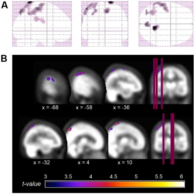
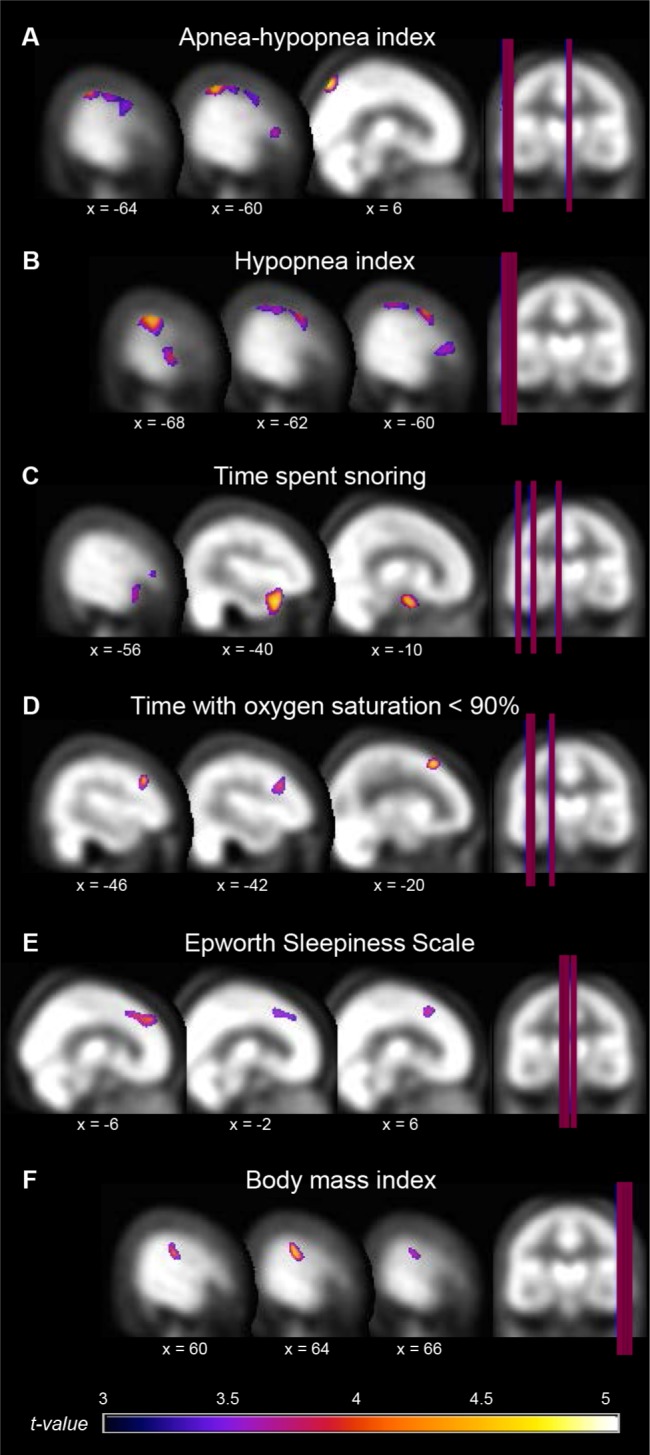
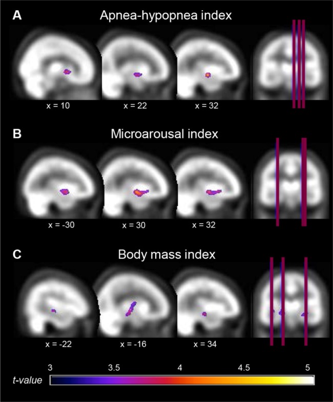
Results: Whereas no rCBF change was observed in mild and moderate groups, participants with severe OSA had reduced rCBF compared to controls in the left parietal lobules, left precentral gyrus, bilateral postcentral gyri, and right precuneus. Reduced rCBF in these regions and in areas of the bilateral frontal and left temporal cortex was associated with more hypopneas, snoring, hypoxemia, and sleepiness. Higher apnea, microarousal, and body mass indexes were correlated to increased rCBF in the basal ganglia, insula, and limbic system.
Conclusions: While older individuals with severe obstructive sleep apnea (OSA) had hypoperfusion in the sensorimotor and parietal areas, respiratory variables and subjective sleepiness were correlated with extended regions of hypoperfusion in the lateral cortex. Interestingly, OSA severity, sleep fragmentation, and obesity correlated with increased perfusion in subcortical and medial cortical regions. Anomalies with such a distribution could result in cognitive deficits and reflect impaired vascular regulation, altered neuronal integrity, and/or undergoing neurodegenerative processes.
Keywords: SPECT; aging; cerebral perfusion; neuroimaging; obstructive sleep apnea; regional cerebral blood flow; snoring.
© 2015 Associated Professional Sleep Societies, LLC.
Figures
Comment in
-
Altered Resting Cerebral Blood Flow in Obstructive Sleep Apnea: A Helpful Change or Not?Sleep. 2015 Sep 1;38(9):1345-7. doi: 10.5665/sleep.4962. Sleep. 2015. PMID: 26285008 Free PMC article. No abstract available.
References
-
- American Academy of Sleep Medicine Task Force. Sleep-related breathing disorders in adults: recommendations for syndrome definition and measurement techniques in clinical research. The Report of an American Academy of Sleep Medicine Task Force. Sleep. 1999;22:667–89. - PubMed
-
- Malhotra A, White DP. Obstructive sleep apnoea. Lancet. 2002;20:237–45. - PubMed
-
- Franklin KA. Cerebral haemodynamics in obstructive sleep apnoea and Cheyne-Stokes respiration. Sleep Med Rev. 2002;6:429–41. - PubMed
-
- Valipour A, McGown AD, Makker H, O'Sullivan C, Spiro SG. Some factors affecting cerebral tissue saturation during obstructive sleep apnoea. Eur Respir J. 2002;20:444–50. - PubMed
Publication types
MeSH terms
Grants and funding
LinkOut - more resources
Full Text Sources
Other Literature Sources
Medical
