. 2015 Jun;23(6):1003-1021.
doi: 10.1038/mt.2015.40. Epub 2015 Mar 12.

Action of obestatin in skeletal muscle repair: stem cell expansion, muscle growth, and microenvironment remodeling

Affiliations

Action of obestatin in skeletal muscle repair: stem cell expansion, muscle growth, and microenvironment remodeling

Uxía Gurriarán-Rodríguez et al. Mol Ther. 2015 Jun.

Abstract

The development of therapeutic strategies for skeletal muscle diseases, such as physical injuries and myopathies, depends on the knowledge of regulatory signals that control the myogenic process. The obestatin/GPR39 system operates as an autocrine signal in the regulation of skeletal myogenesis. Using a mouse model of skeletal muscle regeneration after injury and several cellular strategies, we explored the potential use of obestatin as a therapeutic agent for the treatment of trauma-induced muscle injuries. Our results evidenced that the overexpression of the preproghrelin, and thus obestatin, and GPR39 in skeletal muscle increased regeneration after muscle injury. More importantly, the intramuscular injection of obestatin significantly enhanced muscle regeneration by simulating satellite stem cell expansion as well as myofiber hypertrophy through a kinase hierarchy. Added to the myogenic action, the obestatin administration resulted in an increased expression of vascular endothelial growth factor (VEGF)/vascular endothelial growth factor receptor 2 (VEGFR2) and the consequent microvascularization, with no effect on collagen deposition in skeletal muscle. Furthermore, the potential inhibition of myostatin during obestatin treatment might contribute to its myogenic action improving muscle growth and regeneration. Overall, our data demonstrate successful improvement of muscle regeneration, indicating obestatin is a potential therapeutic agent for skeletal muscle injury and would benefit other myopathies related to muscle regeneration.


Figures
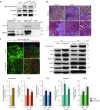
Figure 1
Figure 1
Ectopic expression of preproghrelin enhances muscle regeneration. (a) Upper panel, immunoblot analysis revealed comparable levels of pERK1/2 (T202/Y204) and pAkt (S473) under HA-obestatin stimulation (5 nmol/l, 10 minutes) to obestatin-treated C2C12 myoblast cells (5 nmol/l, 10 minutes). Lower panel, immunoprecipitation analyses using GFP coupled beads demonstrated that GPR39-GFP binds obestatin-HA in C2C12 myoblast cells while it does not bind GFP alone. (b) Representative histology sections of transfected muscle 96 hours and 168 hours after electroporation with CMV plasmid in TA. (c) Left panel, fluorescence microscopy of GFP (pcDNA 3.1-SNAP-GFP, transfection marker) and immunofluorescence analysis of cryosections stained with specific antibodies reactive to laminin and preproghrelin 168 hours after electroporation with the CMV-preproghrelin plasmid in TA. Right panel, representative immunoblots of the mean values from each group of the expression of Pax-7, MyoD, myogenin, and eMHC at 96 and 168 hours after ectopic expression of preproghrelin. Bottom panel, immunoblot analysis of the expression of Pax-7, MyoD, myogenin, and MHC at 96 and 168 hours after ectopic expression of preproghrelin. Protein levels were expressed as fold of control obtained from electroporation of CMV plasmid group (n = 5 per group). Data were expressed as mean ± SEM obtained from intensity scans. *P < 0.05 versus control values.
Figure 2
Figure 2
Ectopic expression of GPR39 or preproghrelin/GPR39 enhances muscle regeneration. (a) Left panel, fluorescence microscopy of GFP (pcDNA 3.1-SNAP-GFP, transfection marker) and immunofluorescence analysis of cryosections stained with antibodies reactive to laminin and GPR39 168 hours after electroporation with the CMV-GPR39 plasmid. Middle panel, representative immunoblots of the mean values from each group of the expression of Pax-7, MyoD, myogenin, and eMHC at 96 and 168 hours after ectopic expression of GPR39. Right panel, immunoblot analysis of the expression of Pax-7, MyoD, myogenin, and MHC at 96 and 168 hours after ectopic expression of GPR39. (b) Left panel, fluorescence microscopy of GFP (pcDNA 3.1-SNAP-GFP, transfection marker) and immunofluorescence analysis of the cryosections stained with antibodies reactive to laminin, preproghrelin or GPR39 168 hours after electroporation with both CMV-GPR39 and CMV-preproghrelin plasmids. Right panel, representative immunoblots of the mean values from each group of the expression of Pax-7, MyoD, myogenin, and MHC at 96 and 168 hours after ectopic expression of GPR39. Bottom panel, immunoblot analysis of the expression of Pax-7, MyoD, myogenin, and eMHC at 96 and 168 hours after ectopic expression of GPR39 and preproghrelin. For a and b, protein levels were expressed as fold of control obtained from electroporation of CMV plasmid group (n = 5 per group). Data were expressed as mean ± SEM obtained from intensity scans. *P < 0.05 versus control values.
Figure 3
Figure 3
Intramuscular obestatin administration enhances muscle regeneration. (a) Upper panel, immunofluorescence analysis of the obestatin dispersion in the distal zone from the injection site (obestatin-HA: 300 nmol/kg body weight; 30 minutes after administration). Lower panel, immunofluorescence analysis of pAkt (S473) of distal cross-sections of obestatin-HA-injected TA, contralateral TA and control TA muscles. Cryosections were stained with antibodies reactive to HA-tag, laminin, pAkt (S473), and counterstained with DAPI 30 minutes after injection of HA-obestatin. (b) Representative images of hematoxylin/eosin-stained sections of resting (control) and freeze-injured TA muscles after intramuscular injection of obestatin (300 nmol/kg body weight/24 hours during 5 days; n = 5 per time point) or vehicle (PBS) at 48, 96, 168, and 240 hours following injury. (c) Effect of intramuscular injection of obestatin (300 nmol/kg body weight/24 hours) on regenerating myofiber (centrally located nuclei) cross-section area at 96 and 168 hours following injury. (d) Cross-sectional area measurements of freeze-injured TA muscles after intramuscular injection of obestatin (300 nmol/kg body weight/24 hours during 5 days; n = 5 per group) at 240 hours following injury. (e) Representative immunoblots of the mean values from each group of the expression of Pax-7, MyoD, myogenin, and eMHC in freeze-injured TA muscles after intramuscular injection of obestatin or vehicle (PBS) at 48, 96, 168, and 240 hours following injury. (e–i) Immunoblot analysis of the expression of Pax-7 (f), MyoD (g), myogenin (h), and eMHC (i) in resting (control) and freeze-injured TA muscles after intramuscular injection of obestatin (300 nmol/kg body weight/24 hours during 5 days; n = 5 per time point) or vehicle (PBS) at 48, 96, 168, and 240 hours following injury. Protein levels were expressed as fold of control (n = 5/group). Data were expressed as mean ± SEM obtained from intensity scans. *,#P < 0.05 versus control values. (j) Representative immunofluorescence images of eMHC in freeze-injured TA muscles after intramuscular injection of obestatin (300 nmol/kg body weight/24 hours during 5 days; n = 5 per time point) or vehicle (PBS) at 96 hours following injury. (k) Serum CK levels at 96 hours postinjury (n = 15 per group). Results are * P < 0.05 versus control values.
Figure 4
Figure 4
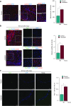
Obestatin activates SCs and increases the number of nuclei per muscle fiber. (a) Average number of myonuclei per 100 TA muscle fibers after intramuscular injection of obestatin (300 nmol/kg body weight/24 hours during 5 days; n = 5 per group) or control (PBS) at 240 hours following injury (n = 5 per group). (b) Sample section for Pax7 and laminin in freeze-injured TA muscle after intramuscular injection of obestatin (300 nmol/kg body weight/24 hours during 5 days; n = 5 per group) or control (PBS) at 96 hours following injury (left panel). Colocalization of Pax7 and DAPI staining from boxed region is shown in the middle panel. The analysis of the relative satellite cell density (ratio of satellite cell number to the cross-section area and normalized to the ratio in control muscles) is shown at 96 hours following injury. Results were calculated from the average values of three muscle sections per mouse, and five mice were analyzed (right panel). (c) Left panel, immunofluorescence analysis of Pax7 positive satellite cells (Pax7+) in myofibers isolated from the neighboring area of the injured zone of the TA muscle after intramuscular injection of obestatin (300 nmol/kg body weight/24 hours during 5 days; n = 5 per group) or control (PBS) at 96 hours following injury. Right panel, Pax7+ cells were enumerated and normalized against myofiber length (µm). Results were calculated from average values of 12 isolated myofibers per mouse, and 5 mice were analyzed per group (n = 60 per group). Data were expressed as mean ± SEM. *P < 0.05 versus control values.
Figure 5
Figure 5
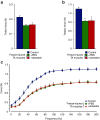
Effect of obestatin administration on muscle force. Obestatin or PBS (vehicle) was administrated via intramuscular injection in freeze-injured TA muscle (300 nmol/kg body weight/24 hours during 5 days). Uninjured TA muscles were taken as control to establish that the damage area was large enough to decrease force (n = 6 per group). Muscle force measurements were assessed after an interval of 5 days after the last dose. (a) Effect of intramuscular injection of obestatin on twitch force at 240 hours following injury. (b) Effect of intramuscular injection of obestatin on tetanic force at 240 hours following injury. (c) Force–frequency curve of TA muscles in control and obestatin- or PBS-treated groups. Data in ac were expressed as mean ± SEM. *P < 0.05 versus control values.
Figure 6
Figure 6
Obestatin regulates the regenerative microenvironment responsible for stimulating and coordinating skeletal muscle repair. Effect of intramuscular injection obestatin (300 nmol/kg body weight/24 hours during 5 days; n = 5 per group) or control (PBS) in freeze-injured TA muscle on Ki67 (a), Cyclin D1 (b), VEGF (c), VEGF-R2 (d), average number of isolectin positive vessels (isolectin+) (e), Col1a2 (f), and myostatin (g) at 96 or 240 hours following injury. Analysis of the expression of Ki67, Cyclin D1, VEGF, VEGF-R2, Col1a2 and myostatin were performed by immunoblot and protein levels were expressed as fold of control (n = 5 per group). Immunoblots are representative of mean values from each group. Data were expressed as mean ± SEM obtained from intensity scans. Isolectin positive vessels were analyzed by immunofluorescence of cryosections stained with isolectin, laminin, and counterstained with DAPI. Representative images of the mean values from each group are shown in the upper panel from e. Representative images of the histological analysis from sections stained with Heidenhain's AZAN trichrome staining are shown in the bottom panel from f. *P < 0.05 versus control values.
Figure 7
Figure 7
Obestatin effect on cell proliferation. (a) Dose–response effect of obestatin (0.01–100 nmol/l) on C2C12 myoblast cell proliferation (48 hours, n = 5). Co: cells initially seeded. Control: cells maintained 48 hours in GM. (b) Immunoblot analysis of Ki67, p57, p21, myogenin, and MHC expression in the course of C2C12 myogenesis under DM or DM + obestatin (5 nmol/l). Protein level was expressed as fold of control cells in GM (n = 3). Immunoblots are representative of the mean value. Data were expressed as mean ± SEM obtained from intensity scans. *P < 0.05 versus control values.
Figure 8
Figure 8
The hypertrophic response to obestatin is not associated with the addition of new myonuclei via proliferation and further fusion. (a) Left panel, immunofluorescence detection of MHC and DAPI in C2C12 myotube cells under DM (control) or DM + obestatin (5 nmol/l) at the 7-day point after stimulation. Right panel, the myotube area (µm2) and the number of nuclei within individual myotubes (at least two nuclei) were evaluated. (b) Left panel, immunofluorescence detection of MHC and DAPI in C2C12 myotube cells under DM (control) or DM + obestatin (5 nmol/l; 3 days of differentiation) at the 7-day point after stimulation. Right panel, the myotube area (µm2) and the number of nuclei within individual myotubes were evaluated. (c) Left panel, immunofluorescence detection of MHC and DAPI in C2C12 myotube cells at 7-day point after stimulation under DM (control) or DM + obestatin (5 nmol/l) + AraC (50 µmol/l) applied at day 3 of differentiation. Right panel, the myotube area (µm2) and the number of nuclei within individual myotubes (at least two nuclei) were evaluated. (d) Immunoblot analysis of myogenin, and MHC expression in C2C12 myotubes at the 7-day point after stimulation under AraC treatment as indicated in c. Immunoblots are representative of mean values from each group. Data were expressed as mean ± SEM obtained from intensity scans. *P < 0.05 versus control values.
Figure 9
Figure 9
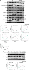
Obestatin-activated intercellular networks involved during myogenic program in C2C12 cells. (a) Immunoblot analysis of pAkt (S473), pERK1/2 (T202/Y204), pp38 (T180/Y182), pS6K1 (S6371), pFoxO1 (T24)/FoxO3a (T32), pCAMKII (T286), and pc-Jun (S63) in the course of C2C12 myogenesis under DM or DM + obestatin (5 nmol/l). (b) Immunoblot analysis of pIGFR (Y1316) and pIRS1 (S636/639) in the course of C2C12 myogenesis under DM or DM + obestatin (5 nmol/l). For a and b, protein level was expressed as fold of control cells in GM (n = 5). Immunoblots are representative of the mean value. Data were expressed as mean ± SEM obtained from intensity scans. *P < 0.05 versus control values.
Figure 10
Figure 10
Obestatin-activated intercellular networks involved during myogenic program in TA muscle 96 hours following injury. (a) Immunoblot analysis of pAkt (S473), pERK1/2 (T202/Y204), pp38 (T180/Y182), pS6K1 (S6371), pFoxO1 (T24)/FoxO3a (T32), pCAMKII (T286), and, pc-Jun (S63) in freeze-injured TA muscles after intramuscular injection of obestatin (300 nmol/kg body weight/24 hours during 5 days; n = 5) or vehicle (PBS) at 96 hours following injury. (b) Immunoblot analysis of pIGFR (Y1316) and pIRS1 (S636/639) in freeze-injured TA muscles after intramuscular injection of obestatin (300 nmol/kg body weight/24 hours during 5 days; n = 5) or vehicle (PBS) at 96 hours following injury. For a and b, protein level was expressed as fold of control TA muscle (n = 5 per group). Immunoblots are representative of the mean value. Data were expressed as mean ± SEM obtained from intensity scans. *P < 0.05 versus control values.

Similar articles

Cited by

References

    1. Yin, H, Price, F and Rudnicki, MA (2013). Satellite cells and the muscle stem cell niche. Physiol Rev 93: 23–67. - PMC - PubMed
    1. Tidball, JG and Villalta, SA (2010). Regulatory interactions between muscle and the immune system during muscle regeneration. Am J Physiol Regul Integr Comp Physiol 298: R1173–R1187. - PMC - PubMed
    1. Chazaud, B, Brigitte, M, Yacoub-Youssef, H, Arnold, L, Gherardi, R, Sonnet, C et al. (2009). Dual and beneficial roles of macrophages during skeletal muscle regeneration. Exerc Sport Sci Rev 37: 18–22. - PubMed
    1. Murray, PJ and Wynn, TA (2011). Protective and pathogenic functions of macrophage subsets. Nat Rev Immunol 11: 723–737. - PMC - PubMed
    1. Tatsumi, R, Anderson, JE, Nevoret, CJ, Halevy, O and Allen, RE (1998). HGF/SF is present in normal adult skeletal muscle and is capable of activating satellite cells. Dev Biol 194: 114–128. - PubMed

Publication types

MeSH terms

Substances