Reduced fetal cerebral oxygen consumption is associated with smaller brain size in fetuses with congenital heart disease
- PMID: 25762062
- PMCID: PMC4398654
- DOI: 10.1161/CIRCULATIONAHA.114.013051
Reduced fetal cerebral oxygen consumption is associated with smaller brain size in fetuses with congenital heart disease
Abstract
Background: Fetal hypoxia has been implicated in the abnormal brain development seen in newborns with congenital heart disease (CHD). New magnetic resonance imaging technology now offers the potential to investigate the relationship between fetal hemodynamics and brain dysmaturation.
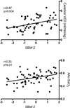
Methods and results: We measured fetal brain size, oxygen saturation, and blood flow in the major vessels of the fetal circulation in 30 late-gestation fetuses with CHD and 30 normal controls using phase-contrast magnetic resonance imaging and T2 mapping. Fetal hemodynamic parameters were calculated from a combination of magnetic resonance imaging flow and oximetry data and fetal hemoglobin concentrations estimated from population averages. In fetuses with CHD, reductions in umbilical vein oxygen content (P<0.001) and failure of the normal streaming of oxygenated blood from the placenta to the ascending aorta were associated with a mean reduction in ascending aortic saturation of 10% (P<0.001), whereas cerebral blood flow and cerebral oxygen extraction were no different from those in controls. This accounted for the mean 15% reduction in cerebral oxygen delivery (P=0.08) and 32% reduction cerebral Vo2 in CHD fetuses (P<0.001), which were associated with a 13% reduction in fetal brain volume (P<0.001). Fetal brain size correlated with ascending aortic oxygen saturation and cerebral Vo2 (r=0.37, P=0.004).
Conclusions: This study supports a direct link between reduced cerebral oxygenation and impaired brain growth in fetuses with CHD and raises the possibility that in utero brain development could be improved with maternal oxygen therapy.
Keywords: brain; heart diseases; hemodynamics; magnetic resonance imaging; pediatrics.
© 2015 American Heart Association, Inc.
Figures


Comment in
-
The path forward is to look backward in time: fetal physiology: the new frontier in managing infants with congenital heart defects.Circulation. 2015 Apr 14;131(15):1307-9. doi: 10.1161/CIRCULATIONAHA.115.016024. Epub 2015 Mar 11. Circulation. 2015. PMID: 25762063 Free PMC article. No abstract available.
-
Letter by Rudolph Regarding Article, "Reduced Fetal Cerebral Oxygen Consumption Is Associated With Smaller Brain Size in Fetuses With Congenital Heart Disease".Circulation. 2016 Jan 5;133(1):e7. doi: 10.1161/CIRCULATIONAHA.115.018348. Circulation. 2016. PMID: 26719395 No abstract available.
-
Response to Letter Regarding Article, "Reduced Fetal Cerebral Oxygen Consumption Is Associated With Smaller Brain Size in Fetuses With Congenital Heart Disease".Circulation. 2016 Jan 5;133(1):e8. doi: 10.1161/CIRCULATIONAHA.115.018748. Circulation. 2016. PMID: 26719396 No abstract available.
References
-
- Limperopoulos C, Tworetzky W, McElhinney DB, Newburger JW, Brown DW, Robertson RL, Guizard N, McGrath E, Geva J, Annese D, Dunbar-Masterson C, Trainor B, Laussen PC, du Plessis AJ. Brain Volume and Metabolism in Fetuses with Congenital Heart Disease: Evaluation with Quantitative Magnetic Resonance Imaging and Spectroscopy. Circulation. 2010;121:26–33. - PMC - PubMed
-
- Miller SP, McQuillan PS, Hamrick S, Xu D, Glidden DV, Charlton N, Karl T, Azakie A, Ferriero DM, Barkovich AJ, Vigneron DB. Abnormal Brain Development in Newborns with Congenital Heart Disease. N Eng J Med. 2007;357:1928–1938. - PubMed
-
- Beca J, Gunn JK, Coleman L, Hope A, Reed PW, Hunt, Finucane K, Brizard C, Dance B, Shekerdemian LS. New white matter brain injury after infant heart surgery is associated with diagnostic group and the use of circulatory arrest. Circulation. 2013;127:971–979. - PubMed
-
- Donofrio MT, Bremer YA, Schieken RM, Gennings C, Morton LD, Eidem BW, Cetta F, Falkensammer CB, Huhta JC, Kleinman CS. Autoregulation of cerebral blood flow in fetuses with congenital heart disease: the brain sparing effect. Pediatr Cardiol. 2003;24:436–443. - PubMed
Publication types
MeSH terms
Grants and funding
LinkOut - more resources
Full Text Sources
Other Literature Sources
Medical

