. 2015 Mar 24;112(12):3698-703.
doi: 10.1073/pnas.1502960112. Epub 2015 Mar 11.

The Fe-S cluster-containing NEET proteins mitoNEET and NAF-1 as chemotherapeutic targets in breast cancer

Affiliations

The Fe-S cluster-containing NEET proteins mitoNEET and NAF-1 as chemotherapeutic targets in breast cancer

Fang Bai et al. Proc Natl Acad Sci U S A. .

Abstract

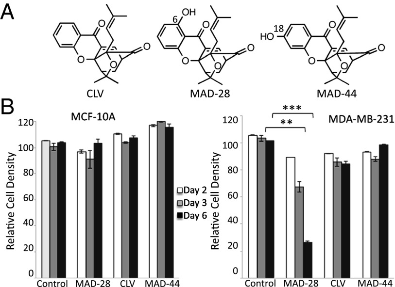
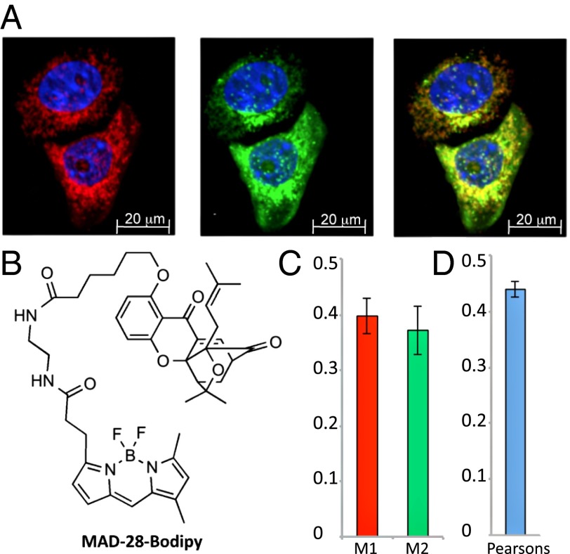
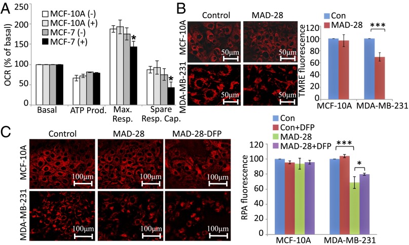
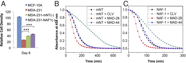
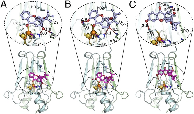
Identification of novel drug targets and chemotherapeutic agents is a high priority in the fight against cancer. Here, we report that MAD-28, a designed cluvenone (CLV) derivative, binds to and destabilizes two members of a unique class of mitochondrial and endoplasmic reticulum (ER) 2Fe-2S proteins, mitoNEET (mNT) and nutrient-deprivation autophagy factor-1 (NAF-1), recently implicated in cancer cell proliferation. Docking analysis of MAD-28 to mNT/NAF-1 revealed that in contrast to CLV, which formed a hydrogen bond network that stabilized the 2Fe-2S clusters of these proteins, MAD-28 broke the coordinative bond between the His ligand and the cluster's Fe of mNT/NAF-1. Analysis of MAD-28 performed with control (Michigan Cancer Foundation; MCF-10A) and malignant (M.D. Anderson-metastatic breast; MDA-MB-231 or MCF-7) human epithelial breast cells revealed that MAD-28 had a high specificity in the selective killing of cancer cells, without any apparent effects on normal breast cells. MAD-28 was found to target the mitochondria of cancer cells and displayed a surprising similarity in its effects to the effects of mNT/NAF-1 shRNA suppression in cancer cells, causing a decrease in respiration and mitochondrial membrane potential, as well as an increase in mitochondrial iron content and glycolysis. As expected, if the NEET proteins are targets of MAD-28, cancer cells with suppressed levels of NAF-1 or mNT were less susceptible to the drug. Taken together, our results suggest that NEET proteins are a novel class of drug targets in the chemotherapeutic treatment of breast cancer, and that MAD-28 can now be used as a template for rational drug design for NEET Fe-S cluster-destabilizing anticancer drugs.

Keywords: NEET proteins; iron-sulfur proteins; mitocan; mitochondria; rational drug design.


Conflict of interest statement

The authors declare no conflict of interest.

Figures

Fig. 1.
Fig. 1.
Structure and selective cytotoxicity of CLV, MAD-28, and MAD-44. (A) Structures of CLV, MAD-28, and MAD-44. The chemical synthesis of MAD-28 and MAD-44 is described in SI Appendix, Fig. S1B. (B) Effect of MAD-28, CLV, and MAD-44 on cell survival of control noncancerous human breast cells MCF-10A (Left) or human epithelial breast cancer cells MDA-MB-231 (Right). Alamar blue cell viability measurements were performed on cells at 2, 3, and 6 d following application of 1.25 μM MAD-28, CLV, or MAD-44. The results show that the three different compounds had no significant effect on the viability of control cells and that MDA-28 had a potent and selective killing effect on cancer cells. Percentage of cell survival was determined based on cells treated with DMSO. Error bars represent SD obtained from three individual experiments. **P < 0.01; ***P < 0.001.
Fig. 2.
Fig. 2.
Subcellular localization of a MAD-28–Bodipy conjugate. (A) MDA-MB-231 cells labeled with mouse anti-Tom20 and visualized with Alexa Fluor 594 goat anti-mouse (Left) were then incubated with MAD-28–Bodipy (Middle). (Right) Merged image is shown. Nuclei are shown in blue. (B) Structure of the MAD-28–Bodipy. The synthesis of MAD-28–Bodipy is shown in SI Appendix, Fig. S1C. Colocalization efficiency was measured using ImageJ software and is shown as Manders’ coefficients M1 and M2 (C) and Pearson’s correlation coefficient (D). The average and SD were obtained by analysis of at least eight different images.
Fig. 3.
Fig. 3.
Measurements of mitochondrial function of control MCF-10A and malignant MCF-7 human epithelial breast cells treated or untreated with MAD-28. (A) Measurements of maximal respiration (Max. Resp.) and spare respiratory capacity (Spare Resp. Cap.) of MCF-10A and MCF-7 cells treated with MAD-28 (+) or DMSO (−), showing that MAD-28 causes a selective decrease in the mitochondrial activity of cancer cells. Measurements of mitochondrial respiration shown were performed as described in Materials and Methods using an XF-24 Seahorse apparatus. *P < 0.05. OCR, oxygen consumption rate. (B) Images (Left) and a quantitative BAR graph (Right) showing the effect of MAD-28 on MMP in control MCF-10A and breast cancer MDA-MB-231 cells [TMRE fluorescence is shown as percentage of control (Con)]. MAD-28 is shown to have a specific effect on cancer cells, decreasing their MMP. (C) Images (Left) and a quantitative BAR graph (Right) showing mitochondrial iron levels in MDA-MB-231 and MCF-10A cells treated or untreated with MAD-28 (RPA fluorescence is shown as percentage of control). MAD-28 is shown to have a specific effect on cancer cells, causing an increase in their mitochondrial iron levels. Error bars represent SD obtained from three individual experiments. (Scale bars: B, 50 μm and C, 100 μm.) *P < 0.05; **P < 0.01; ***P < 0.001.
Fig. 4.
Fig. 4.
Effect of MAD-28 on the survival of cells with suppressed levels of mNT or NAF-1 and on the cluster stability of mNT and NAF-1. (A) Survival assays of MCF-10A and MDA-MB-231 cells with suppressed levels of mNT [MDA-MB-231–mNT(−)] or NAF-1 [MDA-MB-231–NAF-1(−)], showing that the killing effect of MAD-28 on cancer cells is attenuated in cells with suppressed expression of the NEET protein mNT or NAF-1. ***P < 0.001. (B and C) 2Fe-2S cluster stability of mNT (B) or NAF-1 (C) in the absence or presence of stoichiometric concentrations of CLV, MAD-28, or MAD-44 monitored by UV-Vis spectroscopy at 458 nm, showing that MAD-28 has a destabilizing effect and CLV has a stabilizing effect on the clusters of mNT or NAF-1.
Fig. 5.
Fig. 5.
CLV, MAD-28, and MAD-44 are different in their binding to and potential effects on the [2Fe-2S] clusters of mNT. The predicted binding modes of CLV (A), MAD-28 (B), and MAD-44 (C) to mNT (PDB ID code 2QH7) are shown. The lower part highlights the docking of the compound on mNT, the upper part expands the region near the docking site, and the hydrogen bonds emphasized by dashed lines. mNT is shown in cartoon representation, where sticks show the critical residues of mNT, spheres represent the [2Fe-2S] cluster, and light gray stick ball models are the compounds. Hydrogen bonds are shown as red dashed lines, and coordination bonds are shown as solid magenta lines. When MAD-28 binds to mNT, a coordination bond between the iron ion and the H87 is broken (a dashed magenta line shows an elongated distance of 2.7 Å), potentially destabilizing the [2Fe-2S] cluster.

References

    1. Green DR, Reed JC. Mitochondria and apoptosis. Science. 1998;281(5381):1309–1312. - PubMed
    1. Biasutto L, Dong LF, Zoratti M, Neuzil J. Mitochondrially targeted anti-cancer agents. Mitochondrion. 2010;10(6):670–681. - PubMed
    1. Fulda S, Galluzzi L, Kroemer G. Targeting mitochondria for cancer therapy. Nat Rev Drug Discov. 2010;9(6):447–464. - PubMed
    1. Neuzil J, et al. Vitamin E analogues as a novel group of mitocans: Anti-cancer agents that act by targeting mitochondria. Mol Aspects Med. 2007;28(5-6):607–645. - PubMed
    1. Chantarasriwong O, Batova A, Chavasiri W, Theodorakis EA. Chemistry and biology of the caged Garcinia xanthones. Chemistry. 2010;16(33):9944–9962. - PMC - PubMed

Publication types