

The role of exciton delocalization in the major photosynthetic light-harvesting antenna of plants
- PMID: 25762317
- PMCID: PMC4375621
- DOI: 10.1016/j.bpj.2015.01.019
The role of exciton delocalization in the major photosynthetic light-harvesting antenna of plants
Abstract
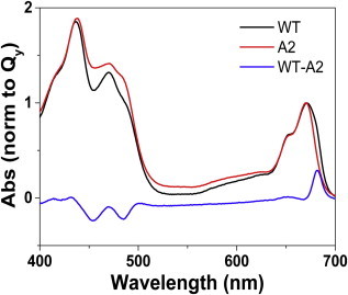
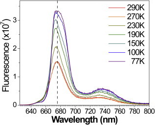
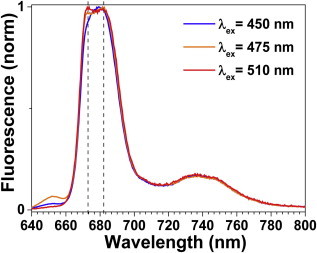
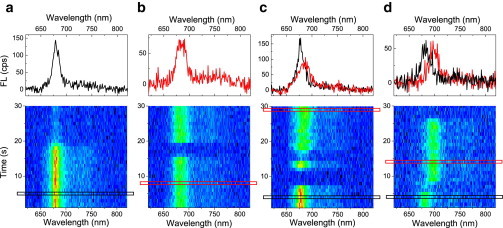
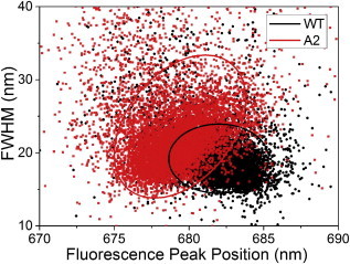
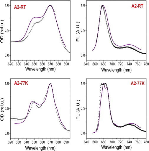
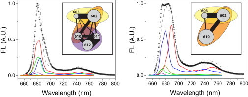
In the major peripheral plant light-harvesting complex LHCII, excitation energy is transferred between chlorophylls along an energetic cascade before it is transmitted further into the photosynthetic assembly to be converted into chemical energy. The efficiency of these energy transfer processes involves a complicated interplay of pigment-protein structural reorganization and protein dynamic disorder, and the system must stay robust within the fluctuating protein environment. The final, lowest energy site has been proposed to exist within a trimeric excitonically coupled chlorophyll (Chl) cluster, comprising Chls a610-a611-a612. We studied an LHCII monomer with a site-specific mutation resulting in the loss of Chls a611and a612, and find that this mutant exhibits two predominant overlapping fluorescence bands. From a combination of bulk measurements, single-molecule fluorescence characterization, and modeling, we propose the two fluorescence bands originate from differing conditions of exciton delocalization and localization realized in the mutant. Disruption of the excitonically coupled terminal emitter Chl trimer results in an increased sensitivity of the excited state energy landscape to the disorder induced by the protein conformations. Consequently, the mutant demonstrates a loss of energy transfer efficiency. On the contrary, in the wild-type complex, the strong resonance coupling and correspondingly high degree of excitation delocalization within the Chls a610-a611-a612 cluster dampens the influence of the environment and ensures optimal communication with neighboring pigments. These results indicate that the terminal emitter trimer is thus an essential design principle for maintaining the efficient light-harvesting function of LHCII in the presence of protein disorder.
Copyright © 2015 Biophysical Society. Published by Elsevier Inc. All rights reserved.
Figures
References
-
- Blankenship R.E. Wiley-Blackwell; Oxford, UK: 2002. Molecular Mechanisms of Photosynthesis.
-
- Fleming G.R., van Grondelle R. The primary steps of photosynthesis. Phys. Today. 1994;47:48–55.
-
- Van Grondelle R., Dekker J.P., Sundstrom V. Energy transfer and trapping in photosynthesis. Biochim. Biophys. Acta Bioenerg. 1994;1187:1–65.
-
- van Grondelle R., Novoderezhkin V.I. Energy transfer in photosynthesis: experimental insights and quantitative models. Phys. Chem. Chem. Phys. 2006;8:793–807. - PubMed
-
- Novoderezhkin V.I., van Grondelle R. Physical origins and models of energy transfer in photosynthetic light-harvesting. Phys. Chem. Chem. Phys. 2010;12:7352–7365. - PubMed
Publication types
MeSH terms
Substances
Grants and funding
LinkOut - more resources
Full Text Sources
Other Literature Sources
