. 2015 Mar 10;108(5):1072-80.
doi: 10.1016/j.bpj.2015.01.026.

Physiological and pharmacological modulation of the embryonic skeletal muscle calcium channel splice variant CaV1.1e

Affiliations

Physiological and pharmacological modulation of the embryonic skeletal muscle calcium channel splice variant CaV1.1e

Bruno Benedetti et al. Biophys J. .

Abstract

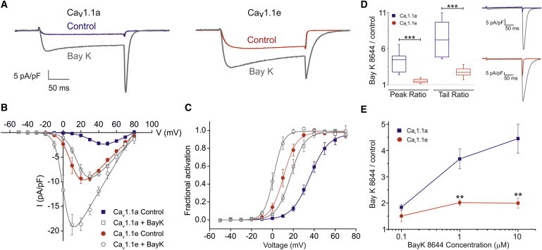
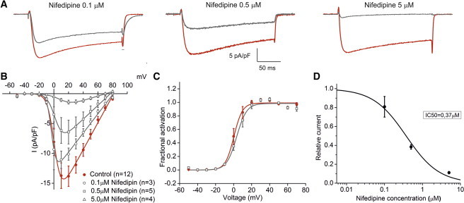
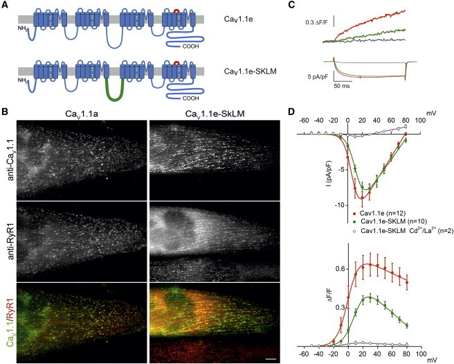
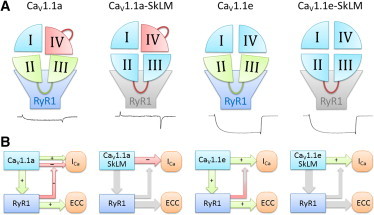
CaV1.1e is the voltage-gated calcium channel splice variant of embryonic skeletal muscle. It differs from the adult CaV1.1a splice variant by the exclusion of exon 29 coding for 19 amino acids in the extracellular loop connecting transmembrane domains IVS3 and IVS4. Like the adult splice variant CaV1.1a, the embryonic CaV1.1e variant functions as voltage sensor in excitation-contraction coupling, but unlike CaV1.1a it also conducts sizable calcium currents. Consequently, physiological or pharmacological modulation of calcium currents may have a greater impact in CaV1.1e expressing muscle cells. Here, we analyzed the effects of L-type current modulators on whole-cell current properties in dysgenic (CaV1.1-null) myotubes reconstituted with either CaV1.1a or CaV1.1e. Furthermore, we examined the physiological current modulation by interactions with the ryanodine receptor using a chimeric CaV1.1e construct in which the cytoplasmic II-III loop, essential for skeletal muscle excitation-contraction coupling, has been replaced with the corresponding but nonfunctional loop from the Musca channel. Whereas the equivalent substitution in CaV1.1a had abolished the calcium currents, substitution of the II-III loop in CaV1.1e did not significantly reduce current amplitudes. This indicates that CaV1.1e is not subject to retrograde coupling with the ryanodine receptor and that the retrograde coupling mechanism in CaV1.1a operates by counteracting the limiting effects of exon 29 inclusion on the current amplitude. Pharmacologically, CaV1.1e behaves like other L-type calcium channels. Its currents are substantially increased by the calcium channel agonist Bay K 8644 and inhibited by the calcium channel blocker nifedipine in a dose-dependent manner. With an IC50 of 0.37 μM for current inhibition by nifedipine, CaV1.1e is a potential drug target for the treatment of myotonic dystrophy. It might block the excessive calcium influx resulting from the aberrant expression of the embryonic splice variant CaV1.1e in the skeletal muscles of myotonic dystrophy patients.


Figures

Figure 1
Figure 1
Bay K 8644 sensitivity of the embryonic CaV1.1e and adult CaV1.1a calcium channel splice variants in skeletal myotubes. (A) Representative patch clamp recordings before and after Bay K 8644 (gray) application from dysgenic myotubes reconstituted with either CaV1.1a (blue) or CaV1.1e (red). Currents (IMax) elicited by depolarizing pulses to peak current potential (VMax), charge carrier 10 mM calcium. (B and C) I/V curves and fractional activation plots indicate that 10 μM Bay K 8644 augments the current densities and left-shifts the voltage-dependence of activation in both CaV1.1a (blue) and CaV1.1e (red). (D) Box plots (left) of the ratio between peak currents before and after application of 10 μM Bay K 8644 recorded at +40 mV test pulses, and of the ratio between tail currents recorded at −80 mV following a +60 mV test pulse. Both ratios are significantly larger in Cav1.1a than in Cav1.1e (p = 0.002). Representative tail currents (right) of Cav1.1a (blue) and Cav1.1e (red) before and after Bay K 8644 application (gray). (E) Relative increase in IMax of CaV1.1a (blue) or CaV1.1e (red) stimulated by three different Bay K 8644 concentrations. The effect of Bay K 8644 is much larger and concentration dependent in the small currents of Cav1.1a compared to the large Cav1.1e currents. To see this figure in color, go online.
Figure 2
Figure 2
Nifedipine block of CaV1.1e calcium currents. (A) Representative calcium current recordings before and after application of 0.1, 0.5, or 5 μM of the LTCC-blocker nifedipine in dysgenic myotubes transfected with GFP-α1S-ΔE29 (CaV1.1e). (B) I/V curves show that nifedipine decreases the current density at all voltages in a dose-dependent manner. (C) Nifedipine (0.5 μM) did not affect the voltage-dependence of current activation. (D) The dose-response curve of nifedipine-block indicates a IC50 of 0.37 μM. To see this figure in color, go online.
Figure 3
Figure 3
CaV1.1e calcium currents are not augmented by retrograde coupling with the RyR1. (A) Domain structure of CaV1.1 depicting the location of the sequence coded by exon 29 (red), which is missing in the embryonic CaV1.1e variant, and the II-III loop that has been swapped with the nonfunctional loop of the Musca channel in CaV1.1e-SkLM (green). (B) Double-immunofluorescence labeling of CaV1.1e variants and the RyR1 in dysgenic myotubes transfected with either CaV1.1e or CaV1.1e-SkLM. Both channels form clusters colocalized with the RyR1. Bar, 10 μm. (C) Representative current traces and fluorescence calcium recordings from myotubes expressing CaV1.1e or CaV1.1e-SkLM. (D) Voltage-dependence of peak current densities and fluorescent calcium transients. CaV1.1e-SkLM (green), compared to CaV1.1e (red), expresses similar size calcium currents but decreased calcium transients, which are fully blocked by Cd2+/La3+ (gray). To see this figure in color, go online.
Figure 4
Figure 4
Gating model explaining the effects of exon 29 and the II-III loop-RyR1 interactions on skeletal muscle calcium currents. (A) In the adult splice variant CaV1.1a the presence of the extracellular loop encoded by exon 29 impedes channel gating (red domain IV). However, interactions of the II-III loop with RyR1 counteract and partially alleviate this inhibition (green), enabling small calcium currents in wild-type CaV1.1a. Without this interaction, as in the CaV1.1a-SkLM mutant, exon 29-containing channels conduct virtually no currents. In the absence of exon 29 in the embryonic splice variant CaV1.1e there is no current inhibition by domain IV. Consequently, there is nothing to alleviate by the II-III loop interactions with RyR1 and its substitution in CaV1.1e-SkLM does not alter the current density. (B) Our current and published data are best explained by a model according to which retrograde coupling with the RyR1 relieves an inhibitory mechanism that exists in CaV1.1a but not in CaV1.1e. Without coupling to the RyR1 the intrinsic inhibitory mechanism affected by exon 29 prevails in CaV1.1a-SkLM. In the absence of this inhibitory mechanism, as in CaV1.1e and CaV1.1e-SkLM, retrograde coupling is ineffective. To see this figure in color, go online.

References

    1. Melzer W., Herrmann-Frank A., Lüttgau H.C. The role of Ca2+ ions in excitation-contraction coupling of skeletal muscle fibres. Biochim. Biophys. Acta. 1995;1241:59–116. - PubMed
    1. Nakai J., Dirksen R.T., Allen P.D. Enhanced dihydropyridine receptor channel activity in the presence of ryanodine receptor. Nature. 1996;380:72–75. - PubMed
    1. Perez-Reyes E., Kim H.S., Birnbaumer L. Induction of calcium currents by the expression of the alpha 1-subunit of the dihydropyridine receptor from skeletal muscle. Nature. 1989;340:233–236. - PubMed
    1. Tuluc P., Molenda N., Jurkat-Rott K. A CaV1.1 Ca2+ channel splice variant with high conductance and voltage-sensitivity alters EC coupling in developing skeletal muscle. Biophys. J. 2009;96:35–44. - PMC - PubMed
    1. Tuluc P., Flucher B.E. Divergent biophysical properties, gating mechanisms, and possible functions of the two skeletal muscle Ca(V)1.1 calcium channel splice variants. J. Muscle Res. Cell Motil. 2011;32:249–256. - PMC - PubMed

Publication types

MeSH terms

Substances

LinkOut - more resources