. 2015 Mar 30;6(9):7136-50.
doi: 10.18632/oncotarget.3044.

Both mTORC1 and mTORC2 are involved in the regulation of cell adhesion

Affiliations

Both mTORC1 and mTORC2 are involved in the regulation of cell adhesion

Long Chen et al. Oncotarget. .

Abstract

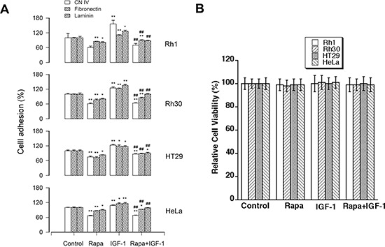
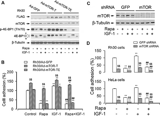
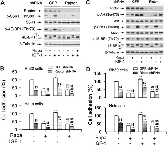
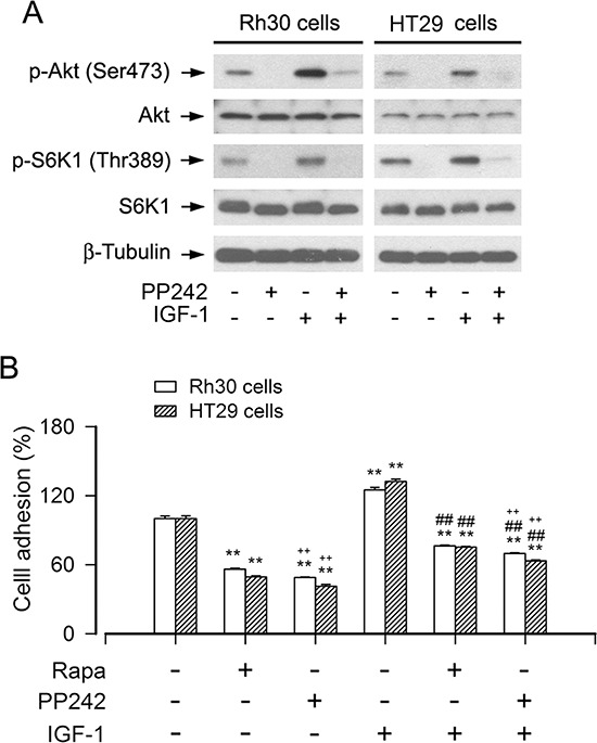
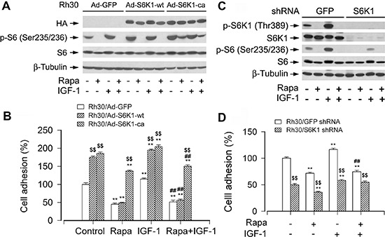
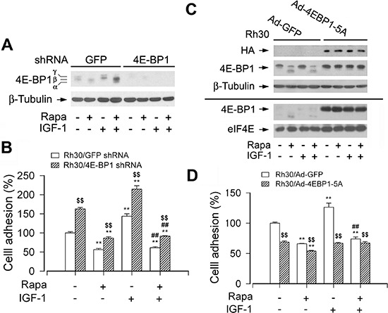
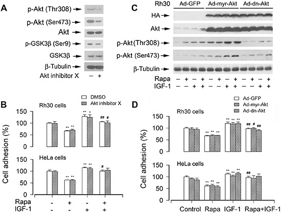
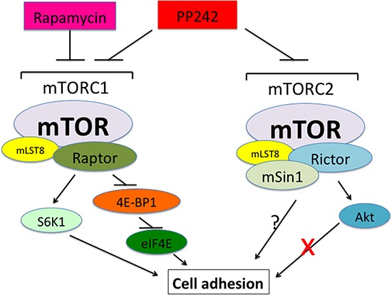
mTOR is a central controller for cell growth/proliferation and survival. Recent studies have shown that mTOR also regulates cell adhesion, yet the underlying mechanism is not known. Here we found that inhibition of mTOR by rapamycin reduced the basal or type I insulin-like growth factor (IGF-1)-stimulated adhesion of cancer cells. Further research revealed that both mTORC1 and mTORC2 were involved in the regulation of cell adhesion, as silencing expression of raptor or rictor inhibited cell adhesion. Also, PP242, an mTORC1/2 kinase inhibitor, inhibited cell adhesion more potently than rapamycin (mTORC1 inhibitor). Of interest, ectopic expression of constitutively active and rapamycin-resistant mutant of p70 kinase 1 (S6K1) or downregulation of eukaryotic initiation factor 4E (eIF4E)-binding protein 1 (4E-BP1) conferred resistance to rapamycin inhibition of cell adhesion, whereas expression of constitutively hypophosphorylated 4E-BP1 (4EBP1-5A) or downregulation of S6K1 suppressed cell adhesion. In contrast, neither genetic manipulation of Akt activity nor pharmacological inhibition of Akt affected cell adhesion. The results suggest that both mTORC1 and mTORC2 are involved in the regulation of cell adhesion; and mTORC1 regulates cell adhesion through S6K1 and 4E-BP1 pathways, but mTORC2 regulates cell adhesion via Akt-independent mechanism.

Keywords: 4E-BP1; Akt; cell adhesion; mTOR; rapamycin.


Conflict of interest statement

CONFLICT OF INTEREST

The authors declare no conflict of interest.

Figures

Figure 1
Figure 1. Rapamycin inhibits the basal or IGF-1-stimulated cell adhesion
Rh30, Rh1, HT29 and HeLa cells were treated with or without rapamycin (Rapa, 100 ng/ml) in the presence or absence of IGF-1 (10 ng/ml) for 1.5 h following pre-incubation with rapamycin for 2 h, respectively. (A) Adherent cells were determined using CN IV-, fibronectin- or laminin-coated cell adhesion assay, and (B) cell viability was evaluated by MTS assay, as described in Materials and Methods. Results are means ± SE (n = 4–12). *P < 0.05, **P < 0.01, difference versus control group. ##P < 0.01, difference versus IGF-1 group.
Figure 2
Figure 2. mTOR kinase activity is essential for cell adhesion
Serum-starved Rh30 and/or HeLa cells, infected with Ad-mTOR-T, Ad-mTOR-TE, or Ad-GFP (for control), or with lentiviral shRNAs to mTOR or GFP, were treated with or without rapamycin (Rapa, 100 ng/ml) for 2 h, followed by stimulation with or without IGF-1 (10 ng/ml) for 1 h. (A and C) Total cell lysates were subjected to Western blotting using indicated antibodies. The blots were probed for β-tubulin as a loading control. Similar results were observed in at least three independent experiments. (B and D) Adherent cells were determined using CN IV-coated cell adhesion assay. (A) Western blot analysis showed stable expression of FLAG-tagged mutants of mTOR in Rh30 cells infected with Ad-mTOR-T and Ad-mTOR-TE, but not in the control cells infected with Ad-GFP. Expression of mTOR-T, but not mTOR-TE or GFP, prevented rapamycin inhibition of the basal or IGF-1-stimulated phosphorylation of 4E-BP1 (Thr70) in Rh30 cells. (B) Ectopic expression of mTOR-T strongly increased cell adhesion and conferred high resistance to rapamycin, whereas expression of mTOR-TE remained sensitive to rapamycin. (C) Lentiviral shRNA to mTOR, but not GFP, downregulated mTOR in Rh30 cells. (D) Downregulation of mTOR inhibited the basal and IGF-1-stimulated adhesion in Rh30 and HeLa cells. Results are means ± SE (n = 12). *P < 0.05, **P < 0.01, difference versus control group. ##P < 0.01, difference versus IGF-1 group. $$P < 0.01, Ad-mTOR-T group versus Ad-GFP group, or mTOR shRNA group versus GFP shRNA group.
Figure 3
Figure 3. Disruption of mTORC1 or mTORC2 suppresses cell adhesion
Serum-starved Rh30 and/or HeLa cells, infected with lentiviral shRNAs to raptor, rictor, or GFP (for control), were treated with or without rapamycin (Rapa, 100 ng/ml) for 2 h, followed by stimulation with or without IGF-1 (10 ng/ml) for 1 h. (A and C) Total cell lysates were subjected to Western blotting using indicated antibodies. The blots were probed for β-tubulin as a loading control. Similar results were observed in at least three independent experiments. (B and D) Adherent cells were determined using CN IV-coated cell adhesion assay. (A) Lentiviral shRNA to raptor, but not GFP, downregulated raptor and prevented the basal and IGF-1-stimulated phosphorylation of S6K1 and 4E-BP1. (B) Downregulation of raptor inhibited the basal and IGF-1-stimulated cell adhesion in Rh30 and HeLa cells. (C) Lentiviral shRNA to rictor, but not GFP, downregulated rictor and inhibited phosphorylation of Akt (Ser473) in Rh30 cells. (D) Downregulation of rictor inhibited the basal and IGF-1-stimulated cell adhesion in Rh30 and HeLa cells. Results are means ± SE (n = 12). **P < 0.01, difference versus control group. ##P < 0.01, difference versus IGF-1 group. $$P < 0.01, raptor shRNA group or rictor shRNA group versus GFP shRNA group.
Figure 4
Figure 4. Inhibition of mTORC1/2 by PP242 potently suppresses the basal or IGF-1-stimulated cell adhesion
Serum-starved Rh30 and HT29 cells were treated with or without PP242 (1 μM) or rapamycin (Rapa, 100 ng/ml) for 2 h, followed by stimulation with or without IGF-1 (10 ng/ml) for 1 h. (A) Total cell lysates were subjected to Western blotting using indicated antibodies, showing that PP242 potently inhibited the basal or IGF-1-stimulated phosphorylation of Akt (Ser473) and S6K1 (Thr389) in Rh30 and HT29 cells. The blots were probed for β-tubulin as a loading control. Similar results were observed in at least three independent experiments. (B) Cell adhesion was determined using CN IV-coated cell adhesion assay, showing that inhibition of mTORC1/2 by PP242 dramatically suppressed the basal and IGF-1-stimulated adhesion in Rh30 and HT29 cells, and the inhibitory effect of PP242 was more potent that of Rapa. Results are means ± SE (n = 6). **P < 0.01, difference versus control group; ##P < 0.01, difference versus IGF-1 group; ++P < 0.01, PP242 group or PP242+IGF-1 group versus Rapa group or Rapa+IGF-1 group.
Figure 5
Figure 5. mTORC1-mediated S6K1 pathway is involved in the regulation of cell adhesion
Serum-starved Rh30 cells, infected with Ad-S6K1-wt, Ad-S6K1-ca, or Ad-GFP (for control), were treated with or without rapamycin (Rapa, 100 ng/ml) for 2 h, followed by stimulation with IGF-1 (10 ng/ml) for 1 h. (A and C) Total cell lysates were subjected to Western blotting using indicated antibodies. The blots were probed for β-tubulin as a loading control. Similar results were observed in at least three independent experiments. (B and D) Adherent cells were determined using CN IV-coated cell adhesion assay. (A) and (B) Rh30 cells expressing S6K1-ca, but not S6K1-wt or GFP, were resistant to rapamycin inhibition of phosphorylation of S6, as well as basal and IGF-1-stimulated cell adhesion. (C) Lentiviral shRNA to S6K1, but not GFP, downregulated S6K1 in Rh30 cells. (D) Downregulation of S6K1 significantly inhibited the basal and IGF-1-stimulated cell adhesion in Rh30 cells. Results are means ± SE (n = 12). **P < 0.01, difference versus control group; ##P < 0.01, difference versus IGF-1 group; $$P < 0.01, Ad-S6K1-wt or Ad-S6K1-ca group versus Ad-GFP group, or S6K1 shRNA group versus GFP shRNA group.
Figure 6
Figure 6. mTORC1-mediated 4E-BP1 pathway is involved in the regulation of cell adhesion
Serum starved Rh30 cells, infected with lentiviral shRNAs to 4E-BP1 or GFP (for control), or with Ad-4EBP1-5A and Ad-GFP, were treated with or without rapamycin (Rapa, 100 ng/ml) for 2 h, followed by stimulation with or without IGF-1 (10 ng/ml) for 1 h. (A and C) Total cell lysates were subjected to Western blotting using indicated antibodies. The blots were probed for β-tubulin as a loading control. Similar results were observed in at least three independent experiments. (B and D) Adherent cells were determined using CN IV-coated cell adhesion assay. (A) Lentiviral shRNA to 4E-BP1, but not GFP, resulted in a significant downregulation of 4E-BP1 in Rh30 cells. (B) Downregulation of 4E-BP1 significantly elevated the basal and IGF-1-stimulated cell adhesion in Rh30 cells. (C) Exposure to rapamycin due to HA-tagged 4E-BP1 in Ad-4EBP1-5A-infected cells failed to change the mobility of 4E-BP1-5A (upper panel). However, using 7-methyl GTP-Sepharose pull-down assay, very striking amount of recombinant 4EBP1-5A was observed to bind to eIF4E, even in the absence of rapamycin (bottom panel). (D) Expression of 4EBP1-5A inhibited the basal and IGF-1-stimulated cell adhesion in Rh30 cells. Results are means ± SE (n = 12). **P < 0.01, difference versus control group; ##P < 0.01, difference versus IGF-1 group; $$P < 0.01, GFP shRNA group versus 4E-BP1 shRNA group, or Ad-4EBP1-5A group versus Ad-GFP group.
Figure 7
Figure 7. Pharmacological inhibition of Akt or ectopic expression of constitutively active or dominant negative Akt does not affect cell adhesion
(A) Serum-starved Rh30 were treated with or without Akt inhibitor X (10 μM) for 2 h, followed by Western blot analysis using indicated antibodies, showing that Akt inhibitor X inhibited the phosphorylation of Akt and its substrate GSK3β in the cells. (B) The adhesion of Rh30 and HeLa cells, treated with or without rapamycin and/or IGF-1 following pre-incubation with or without Akt inhibitor X for 2 h, was not significantly affected. (C and D) Serum starved Rh30 and/or HeLa cells, infected with Ad-myr-Akt, Ad-dn-Akt, or Ad-GFP (for control), were treated with or without rapamycin (Rapa, 100 ng/ml) for 2 h, followed by stimulation with or without IGF-1 (10 ng/ml) for 1 h. (C) Total cell lysates were subjected to Western blotting using indicated antibodies. The blots were probed for β-tubulin as a loading control. Similar results were observed in at least three independent experiments. (D) Adherent cells were determined using CN IV-coated cell adhesion assay, showing that ectopic expression of myr-Akt or dn-Akt did not exhibit an obvious stimulatory or inhibitory effect on cell adhesion in Rh30 and HeLa cells. Results are means ± SE (n = 12). *P < 0.05, **P < 0.01, difference versus control group; #P < 0.01, ##P < 0.01, difference versus IGF-1 group.
Figure 8
Figure 8. A schematic model showing how mTOR regulates cell adhesion
Both mTORC1 and mTORC2 control cell adhesion. mTORC1 regulates cell adhesion through S6K1 and 4E-BP1/eIF4E pathways, but mTORC2 mediates cell adhesion independently of Akt.

Comment in

  • A deut of mTORC1/2 for cell adhesion.
    Chen L, Liu C, Huang S. Chen L, et al. Cell Cycle. 2015;14(8):1131-2. doi: 10.1080/15384101.2015.1022075. Cell Cycle. 2015. PMID: 25789977 Free PMC article. No abstract available.

References

    1. Woodhouse EC, Chuaqui RF, Liotta LA. General mechanisms of metastasis. Cancer. 1997;80:1529–1537. - PubMed
    1. Leber MF, Efferth T. Molecular principles of cancer invasion and metastasis. Int J Oncol. 2009;34:881–895. - PubMed
    1. Cornu M, Albert V, Hall MN. mTOR in aging, metabolism, and cancer. Curr Opin Genet Dev. 2013;23:53–62. - PubMed
    1. Lamming DW, Ye L, Sabatini DM, Baur JA. Rapalogs and mTOR inhibitors as anti-aging therapeutics. J Clin Invest. 2013;123:980–989. - PMC - PubMed
    1. Loewith R, Jacinto E, Wullschleger S, Lorberg A, Crespo JL, Bonenfant D, Oppliger W, Jenoe P, Hall MN. Two TOR complexes, only one of which is rapamycin sensitive, have distinct roles in cell growth control. Mol Cell. 2002;10:457–468. - PubMed

Publication types

MeSH terms

Substances