. 2015 Mar 11;35(10):4190-202.
doi: 10.1523/JNEUROSCI.3592-14.2015.

Removal of perineuronal nets in the medial prefrontal cortex impairs the acquisition and reconsolidation of a cocaine-induced conditioned place preference memory

Affiliations

Removal of perineuronal nets in the medial prefrontal cortex impairs the acquisition and reconsolidation of a cocaine-induced conditioned place preference memory

Megan Slaker et al. J Neurosci. .

Erratum in

  • J Neurosci. 2015 May 27;35(21):8376

Abstract

Pyramidal neurons in the medial prefrontal cortex (mPFC) critically contribute to cocaine-seeking behavior in humans and rodents. Activity of these neurons is significantly modulated by GABAergic, parvalbumin-containing, fast-spiking interneurons, the majority of which are enveloped by specialized structures of extracellular matrix called perineuronal nets (PNNs), which are integral to the maintenance of many types of plasticity. Using a conditioned place preference (CPP) procedure, we found that removal of PNNs primarily from the prelimbic region of the mPFC of adult, male, Sprague Dawley rats impaired the acquisition and reconsolidation of a cocaine-induced CPP memory. This impairment was accompanied by a decrease in the number of c-Fos-positive cells surrounded by PNNs. Following removal of PNNs, the frequency of inhibitory currents in mPFC pyramidal neurons was decreased; but following cocaine-induced CPP, both frequency and amplitude of inhibitory currents were decreased. Our findings suggest that cocaine-induced plasticity is impaired by removal of prelimbic mPFC PNNs and that PNNs may be a therapeutic target for disruption of cocaine CPP memories.

Keywords: cocaine; conditioned place preference; memory; perineuronal net.


Figures

Figure 1.
Figure 1.
Cocaine-associated reward increases WFA/c-Fos double-labeled neurons in the PL mPFC. A, Timeline of experimental procedure. B, Time spent in cocaine-paired chamber on initial preference and test day for CPP. Animals conditioned in a paired manner (n = 8/group) show place preference, but animals conditioned in an unpaired manner (n = 8) do not show preference. C, c-Fos-labeled cells within layers V and VI of the PL mPFC are increased 60 min after confinement to the cocaine-paired, but not saline-paired, chamber in animals trained in a paired manner. c-Fos-labeled cells within layers V and VI of the IL mPFC are increased 60 min after confinement to either the cocaine-paired or saline-paired chamber in animals trained in a paired manner. D, WFA-labeled cells within layers V and VI of the PL mPFC are decreased 60 min after confinement to the cocaine- or saline-paired chamber in animals trained in a paired manner. WFA-labeled cells within layers V and VI of the IL mPFC are decreased only in animals trained in a paired manner and confined to the saline-paired chamber. E, WFA/c-Fos double-labeled neurons within layers V and VI of the PL mPFC, but not the IL mPFC, were increased in animals confined to the cocaine-paired chamber compared with those confined to the saline-paired chamber or to those confined to a pseudo-conditioned chamber. F–H, Representative images from the PL mPFC of pseudo-conditioned (F), saline-paired (G), and cocaine-paired (H) animals. Arrows indicate double-labeled cells for WFA (green) and c-Fos (red). Scale bar, 100 μm. Data are mean ± SEM. *p < 0.05, compared with vehicle-treated animals. $p < 0.05, compared with pseudo-conditioned animals. +p < 0.05, compared with initial preference day.
Figure 2.
Figure 2.
Intra-mPFC cannulae placements. Circles represent the most ventral point of the cannula tract for each animal receiving treatment primarily within the PL mPFC. Squares represent the most ventral location of cannulae tracts for each animal receiving intra-IL treatment. Numbers indicate the distance from bregma in millimeters.
Figure 3.
Figure 3.
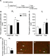
Intra-PL administration of Ch-ABC attenuates acquisition of cocaine-induced CPP. A, Timeline of experimental procedure. B, Time spent in cocaine-paired chamber on initial preference and test day for CPP is shown after pretreatment with intra-PL administration of vehicle (n = 11) or Ch-ABC (n = 13). C, Time spent in cocaine-paired chamber on initial preference and test day for CPP is shown after pretreatment with intra-IL administration of vehicle (n = 5) or Ch-ABC (n = 6). D, Average number of WFA/c-Fos double-labeled neurons in the PL mPFC treated with vehicle (n = 5) and Ch-ABC (n = 5). Rats treated with Ch-ABC had fewer WFA/c-Fos double-labeled neurons than rats treated with vehicle. E, F, Representative images from rats treated with vehicle (E) and Ch-ABC (F) of WFA (green) and c-Fos (red). Arrow indicates double-labeled cells. Scale bar, 100 μm. Data are mean ± SEM. *p < 0.05, compared with vehicle-treated animals. +p < 0.05, compared with initial preference day.
Figure 4.
Figure 4.
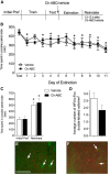
Intra-PL administration of Ch-ABC before extinction does not alter the rate of extinction training or suppress subsequent reinstatement of cocaine-induced CPP. A, Timeline of experimental procedure. B, Time course of extinction rate for vehicle-treated (n = 6) and Ch-ABC-treated (n = 5) rats. Treatment did not alter the rate of extinction. C, Time spent in cocaine-paired chamber on initial preference and reinstatement day (Reinstate) is shown. D, Average number of WFA/c-Fos double-labeled neurons following the reinstatement test was not different between treatment groups. E, F, Representative images from rats treated with vehicle (E) and Ch-ABC (F) of WFA (green) and c-Fos (red). Arrows indicate double-labeled cells. Scale bar, 100 μm. Data are mean ± SEM. +p < 0.05, compared with initial preference day. *p < 0.05, compared with test day.
Figure 5.
Figure 5.
Intra-PL administration of Ch-ABC attenuates reconsolidation of cocaine-induced CPP. A, Timeline of experimental procedure. B, Time spent in cocaine-paired chamber on initial preference day and reinstatement day (Reinstate) is shown for animals (vehicle: n = 18; Ch-ABC: n = 15) receiving treatment 3 d before a reactivation session (React). C, WFA/c-Fos double-labeled neurons within the PL mPFC decreased in animals treated with Ch-ABC compared with animals treated with vehicle. D, E, Representative images from rats treated with vehicle (D) and Ch-ABC (E) of WFA (green) and c-Fos (red). Arrows indicate double-labeled cells. F, Time spent in cocaine-paired chamber on initial preference day and reinstatement day (Reinstate) is shown for animals (vehicle: n = 14; Ch-ABC: n = 16) not receiving a memory reactivation session (No React). G, Average number of WFA/c-Fos double-labeled cells within the PL mPFC was not different between groups. H, I, Representative images from rats treated with vehicle (H) and Ch-ABC (I) of WFA (green) and c-Fos (red). Arrows indicate double-labeled cells. Scale bar, 100 μm. Data are mean ± SEM. *p < 0.05, compared with vehicle treated animals. +p < 0.05, compared with initial preference day.
Figure 6.
Figure 6.
Time course of WFA intensity following Ch-ABC administration. Ch-ABC treatment condition was normalized to control levels for each time point and for both the intensity and cell number analysis. A–D, Three days following treatment, WFA intensity (C) is decreased in Ch-ABC-treated animals compared with vehicle-treated animals (n = 8/group). Representative single-channel confocal micrographs of vehicle-treated (A) and Ch-ABC-treated (B) tissue. D, The relative number of WFA-surrounded cells is decreased following Ch-ABC treatment compared with vehicle treatment (raw cell count: vehicle, 10.31 ± 1.34 cells; Ch-ABC, 3.19 ± 0.78 cells, p < 0.005). E–H, Nine days following treatment, WFA intensity (G) is decreased in Ch-ABC-treated (n = 6) animals compared with vehicle-treated (n = 4) animals. Representative single-channel confocal micrographs of vehicle-treated (E) and Ch-ABC-treated (F) tissue. H, The relative number of WFA-surrounded cells was not different between treatment groups (raw cell count: vehicle, 5.46 ± 0.83; Ch-ABC 3.5 ± 0.57, p = 0.08). I–L, Thirteen days following treatment, WFA intensity (K) is decreased in Ch-ABC-treated (n = 8) compared with vehicle-treated (n = 4) animals. Representative single-channel confocal micrographs of vehicle-treated (I) and Ch-ABC-treated (J) tissue. L, The relative number of WFA-surrounded cells was not different between treatment groups (raw cell count: vehicle, 7.88 ± 1.23; Ch-ABC, 8.31 ± 1.08, p = 0.81). Data are mean ± SEM. *p < 0.05, compared with vehicle-treated animals. Scale bar, 50 μm.
Figure 7.
Figure 7.
Infusion of Ch-ABC into the mPFC increases the excitability of mPFC pyramidal neurons, and mIPSC amplitude and frequency are decreased in mPFC pyramidal neurons following cocaine-induced CPP. A, Example traces of mIPSC recordings from mPFC pyramidal neurons from slices extracted from control (n = 11 rats, 46 neurons), cocaine-induced CPP trained rats (n = 7 rats, 16 neurons), or rats infused with Ch-ABC into the mPFC 9 d earlier (n = 4 rats, 18 neurons). Control slices are pooled data from intra-mPFC vehicle-injected animals and naive animals. Control experiments were interleaved with all other experimental groups. Holding potential, 70 mV. Calibration: 50 pA, 25 ms. B, C, Pyramidal neurons from slices where animals were previously exposed to cocaine-induced CPP training showed significantly attenuated mIPSC amplitudes compared with cells from control slices (Kolmogorov–Smirnov test, p < 0.001). However, mIPSC amplitude was unaltered in brain slices previously microinjected with Ch-ABC (Kolmogorov–Smirnov test, p < 0.51). D, E, Cocaine exposure or infusion of Ch-ABC 9 d before slice preparation significantly reduced mIPSC frequency compared with control slices (ANOVA, p < 0.05). F, Example traces of action potential firing from control rats (n = 4 rats, 10 neurons) and rats infused with Ch-ABC (n = 3 rats, 6 neurons) into the mPFC 9 d before slice preparation at various holding currents: 150, 400, and 650 pA. Calibration: 40 mV, 40 ms. G, Microinjection of Ch-ABC into the mPFC 9 d before slice preparation significantly increased the number of action potentials elicited at various holding currents (two-way ANOVA with Sidak's post hoc, p < 0.01). Data are mean ± SEM. *p < 0.05, compared with control slices.

References

    1. Abel T, Lattal KM. Molecular mechanisms of memory acquisition, consolidation and retrieval. Curr Opin Neurobiol. 2001;11:180–187. doi: 10.1016/S0959-4388(00)00194-X. - DOI - PubMed
    1. Augustine GJ, Kasai H. Bernard Katz, quantal transmitter release and the foundations of presynaptic physiology. J Physiol. 2007;578:623–625. doi: 10.1113/jphysiol.2006.123224. - DOI - PMC - PubMed
    1. Barritt AW, Davies M, Marchand F, Hartley R, Grist J, Yip P, McMahon SB, Bradbury EJ. Chondroitinase ABC promotes sprouting of intact and injured spinal systems after spinal cord injury. J Neurosci. 2006;26:10856–10867. doi: 10.1523/JNEUROSCI.2980-06.2006. - DOI - PMC - PubMed
    1. Brown TE, Forquer MR, Cocking DL, Jansen HT, Harding JW, Sorg BA. Role of matrix metalloproteinases in the acquisition and reconsolidation of cocaine-induced conditioned place preference. Learn Mem. 2007;14:214–223. doi: 10.1101/lm.476207. - DOI - PMC - PubMed
    1. Brückner G, Brauer K, Härtig W, Wolff JR, Rickmann MJ, Derouiche A, Delpech B, Girard N, Oertel WH, Reichenbach A. Perineuronal nets provide a polyanionic, glia-associated form of microenvironment around certain neurons in many parts of the rat brain. Glia. 1993;8:183–200. doi: 10.1002/glia.440080306. - DOI - PubMed

Publication types

MeSH terms