. 2015 Mar 11;35(10):4280-6.
doi: 10.1523/JNEUROSCI.5063-14.2015.

Cockayne syndrome group B (Csb) and group a (Csa) deficiencies predispose to hearing loss and cochlear hair cell degeneration in mice

Affiliations

Cockayne syndrome group B (Csb) and group a (Csa) deficiencies predispose to hearing loss and cochlear hair cell degeneration in mice

A Paul Nagtegaal et al. J Neurosci. .

Abstract

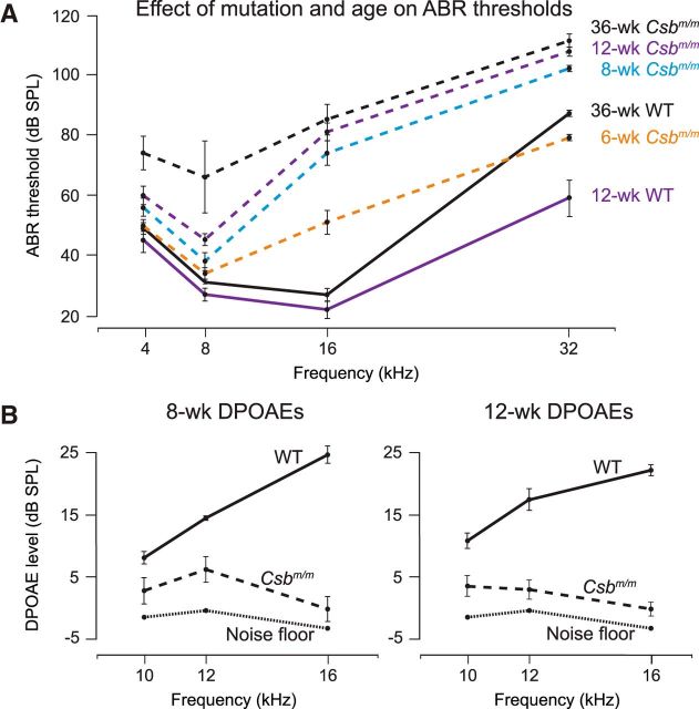
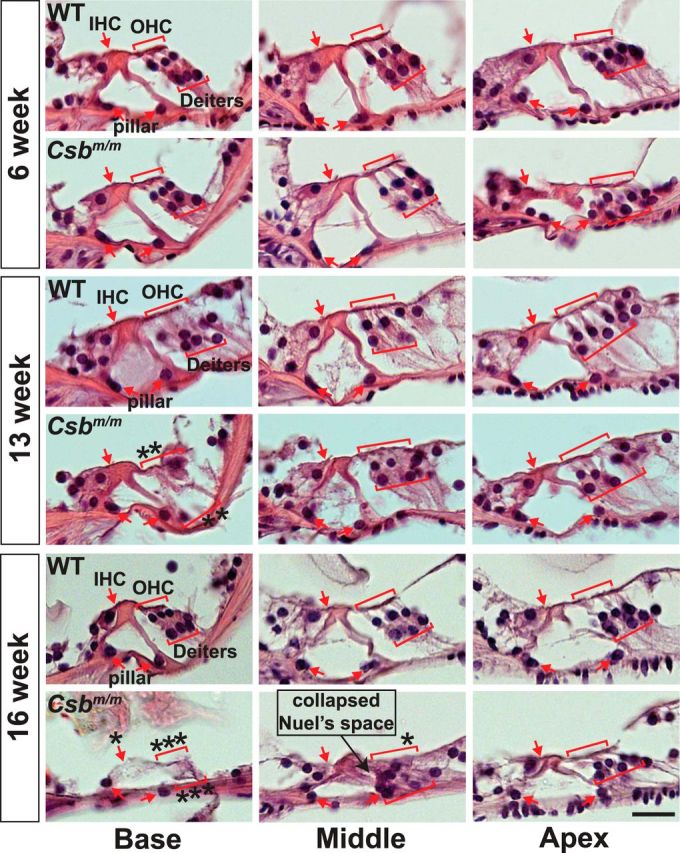
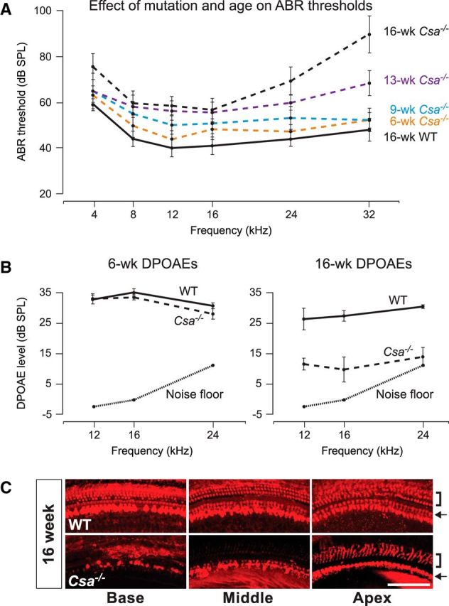
Sensory hair cells in the cochlea, like most neuronal populations that are postmitotic, terminally differentiated, and non-regenerating, depend on robust mechanisms of self-renewal for lifelong survival. We report that hair cell homeostasis requires a specific sub-branch of the DNA damage nucleotide excision repair pathway, termed transcription-coupled repair (TCR). Cockayne syndrome (CS), caused by defects in TCR, is a rare DNA repair disorder with a broad clinical spectrum that includes sensorineural hearing loss. We tested hearing and analyzed the cellular integrity of the organ of Corti in two mouse models of this disease with mutations in the Csb gene (CSB(m/m) mice) and Csa gene (Csa(-/-) mice), respectively. Csb(m/m) and Csa(-/-) mice manifested progressive hearing loss, as measured by an increase in auditory brainstem response thresholds. In contrast to wild-type mice, mutant mice showed reduced or absent otoacoustic emissions, suggesting cochlear outer hair cell impairment. Hearing loss in Csb(m/m) and Csa(-/-) mice correlated with progressive hair cell loss in the base of the organ of Corti, starting between 6 and 13 weeks of age, which increased by 16 weeks of age in a basal-to-apical gradient, with outer hair cells more severely affected than inner hair cells. Our data indicate that the hearing loss observed in CS patients is reproduced in mouse models of this disease. We hypothesize that accumulating DNA damage, secondary to the loss of TCR, contributes to susceptibility to hearing loss.

Keywords: Cockayne syndrome; DNA damage; DNA repair; csb/csa; hair cell; hearing loss.


Figures

Figure 1.
Figure 1.
Csbm/m mice show progressively higher ABR thresholds and reduced DPOAEs in early life. A, ABR thresholds of mice carrying a homozygous mutation in the Csb gene (Csbm/m) and WT controls were measured between 6 and 36 weeks of age. In control mice, measurements at 6 and 8 weeks of age were not significantly different from 12 weeks of age (p = NS for all differences), and thus only 12 week values are shown. The 36-week-old control mice display elevated high-frequency ABR thresholds, as expected in a C57BL/6J strain background (Willott and Erway, 1998); however, thresholds in Csbm/m mice remain significantly higher than those in WT mice at this age (p < 0.01). B, 8- and 12-week-old Csbm/m mice have reduced or absent DPOAEs compared with WT controls. ABR and DPOAE data are shown as the mean ± SEM.
Figure 2.
Figure 2.
Csbm/m mice manifest progressive hair cell loss in a basal-to-apical gradient, with outer hair cells more severely affected than inner hair cells. A, Whole-mount preparations of the organ of Corti from 6-, 13-, and 16-week-old mice carrying a homozygous mutation in the Csb gene (Csbm/m) and WT littermates were stained with an antibody against the hair cell marker MyosinVI. Outer hair cells and inner hair cells were quantified independently along the length of the cochlear duct (base, middle, and apex) for each genotype and age group. Solid line, WT mice; broken line, Csbm/m mice. Error bars represent the SEM. B, Representative pictures from basal, middle, and apical regions are shown. In addition to an apparent basal-to-apical gradient of cell loss, outer hair cells (brackets) are significantly more affected than inner hair cells (arrows) in Csbm/m mice with respect to age. Scale bar, 100 μm.
Figure 3.
Figure 3.
Hematoxylin and eosin-stained paraffin sections through the organ of Corti of Csbm/m mice indicate progressive hair cell and supporting cell loss. Representative sections from each cochlear position obtained at 6, 13, and 16 weeks are pictured. Outer hair cells (OHC), inner hair cells (IHC), Deiters' cells, and pillar cells are indicated in red. OHCs and IHCs are separated by two rows of pillar cells forming the tunnel of Corti. An asterisk indicates a missing cell. Notice the clear deterioration of outer hair cells and Deiters' cells at 16 weeks, leading to the collapse of Nuel's space (formed by outer pillar cells on one side and OHCs/Deiters' cells on the other). Scale bar, 20 μm.
Figure 4.
Figure 4.
Csa−/− mice show progressive elevated ABR thresholds, reduced DPOAEs, and hair cell loss in a basal-to-apical gradient, with outer hair cells more severely affected than inner hair cells, in early life. A, ABR thresholds of mice carrying a homozygous deletion in the Csa gene (Csa−/−) and WT controls were measured between 6 and 16 weeks of age. No significant difference between control mice at any of the ages measured was detected (p = NS for all differences), and thus only 16 week values are shown. Solid line, WT mice; broken line, Csa−/− mice. B, The 6-week-old Csa−/− mice have DPOAEs similar to those of age-matched WT controls, whereas 16-week-old Csa−/− mice have reduced or absent DPOAEs compared with WT controls. C, Representative pictures of 16-week-old Csa−/− and WT cochlear whole-mount preparations stained with an antibody against the hair cell marker parvalbumin. In addition to an apparent basal-to-apical gradient of cell loss, outer hair cells (brackets) are more affected than inner hair cells (arrows) in Csa−/− mice. Scale bar, 100 μm. ABR and DPOAE data are shown as the mean ± SEM.

References

    1. Berg RJ, Rebel H, van der Horst GT, van Kranen HJ, Mullenders LH, van Vloten WA, de Gruijl FR. Impact of global genome repair versus transcription-coupled repair on ultraviolet carcinogenesis in hairless mice. Cancer Res. 2000;60:2858–2863. - PubMed
    1. Coppedè F, Migliore L. DNA repair in premature aging disorders and neurodegeneration. Curr Aging Sci. 2010;3:3–19. doi: 10.2174/1874609811003010003. - DOI - PubMed
    1. Fousteri M, Mullenders LH. Transcription-coupled nucleotide excision repair in mammalian cells: molecular mechanisms and biological effects. Cell Res. 2008;18:73–84. doi: 10.1038/cr.2008.6. - DOI - PubMed
    1. Iwasaki S, Kaga K. Chronological changes of auditory brainstem responses in Cockayne's syndrome. Int J Pediatr Otorhinolaryngol. 1994;30:211–221. doi: 10.1016/0165-5876(94)90062-0. - DOI - PubMed
    1. Jiang H, Talaska AE, Schacht J, Sha SH. Oxidative imbalance in the aging inner ear. Neurobiol Aging. 2007;28:1605–1612. doi: 10.1016/j.neurobiolaging.2006.06.025. - DOI - PMC - PubMed

Publication types

MeSH terms