. 2015 Apr 7;84(14):1473-82.
doi: 10.1212/WNL.0000000000001454. Epub 2015 Mar 11.

Hematopoietic mobilization: Potential biomarker of response to natalizumab in multiple sclerosis

Affiliations

Hematopoietic mobilization: Potential biomarker of response to natalizumab in multiple sclerosis

Miriam Mattoscio et al. Neurology. .

Abstract

Objective: To ascertain the mobilization from the bone marrow and the functional relevance of the increased number of circulating hematopoietic stem and progenitor cells (HSPC) induced by the anti-α-4 integrin antibody natalizumab in patients with multiple sclerosis (MS).

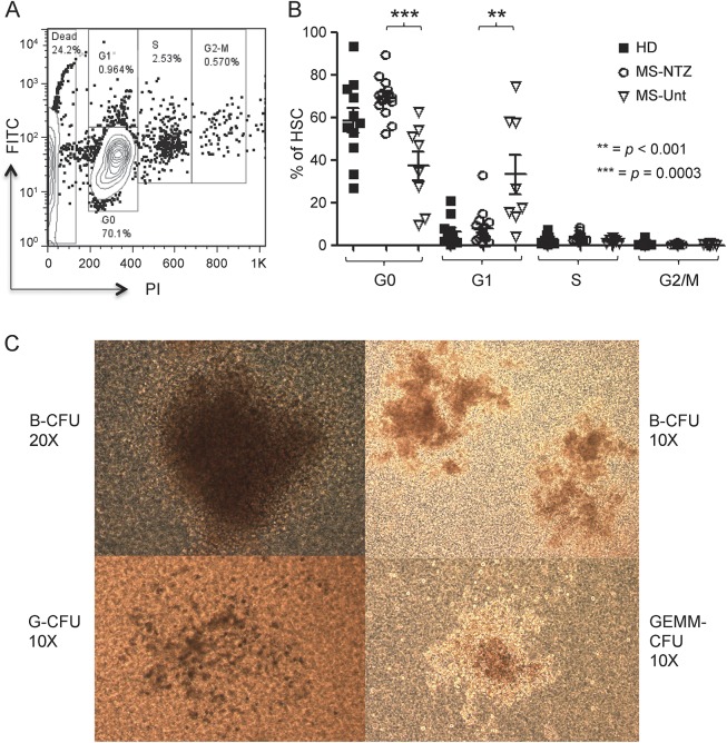
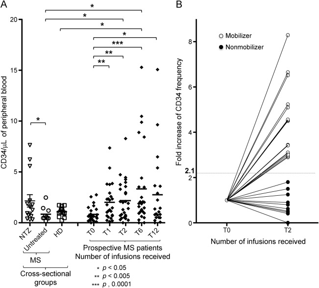
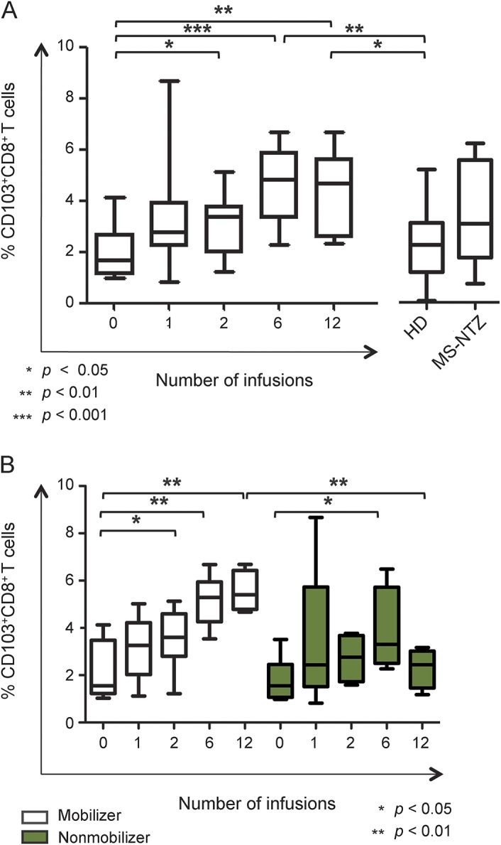
Methods: We evaluated CD45(low)CD34+ HSPC frequency by flow cytometry in blood from 45 natalizumab-treated patients (12 of whom were prospectively followed during the first year of treatment as part of a pilot cohort and 16 prospectively followed for validation), 10 untreated patients with MS, and 24 healthy donors. In the natalizumab-treated group, we also assessed sorted HSPC cell cycle status, T- and B-lymphocyte subpopulation frequencies (n = 29), and HSPC differentiation potential (n = 10).

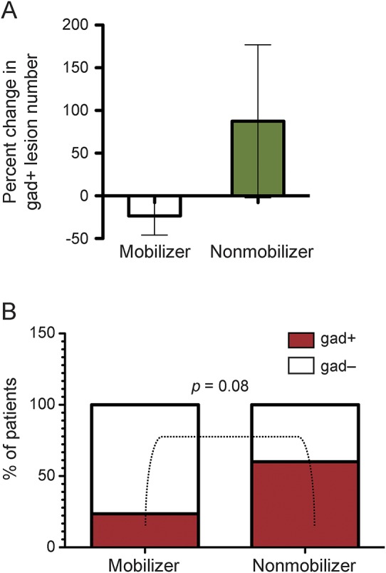
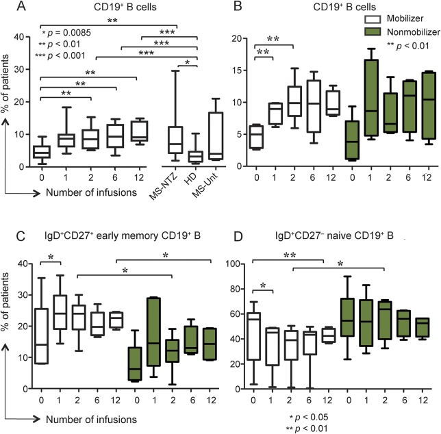
Results: Natalizumab-induced circulating HSPC were predominantly quiescent, suggesting recent mobilization from the bone marrow, and were capable of differentiating ex vivo. Circulating HSPC numbers were significantly increased during natalizumab, but heterogeneously, allowing the stratification of mobilizer and nonmobilizer subgroups. Nonmobilizer status was associated with persistence of disease activity during treatment. The frequency of B cells and CD103+CD8+ regulatory T cells persistently increased, more significantly in mobilizer patients, who also showed a specific naive/memory B-cell profile.

Conclusions: The data suggest that natalizumab-induced circulating HSPC increase is the result of true mobilization from the bone marrow and has clinical and immunologic relevance. HSPC mobilization, associated with clinical remission and increased proportion of circulating B and regulatory T cells, may contribute to the treatment's mode of action; thus, HSPC blood counts could represent an early biomarker of responsiveness to natalizumab.


Figures

Figure 1
Figure 1. Natalizumab-induced circulating hematopoietic stem and progenitor cells are predominantly quiescent and retain full expansion capacity ex vivo
(A) Representative example of flow cytometry acquisition plot shows CD34+ hematopoietic stem and progenitor cells (HSPC) magnetically sorted from peripheral blood mononuclear cells of 1 patient who had received 2 infusions of natalizumab, after labeling with fluorescein isothiocyanate and propidium iodide to measure intracellular protein and DNA content, respectively; G0, G1, S, and G2/M phases of the cell cycle are defined according to the expression of the 2 markers. (B) Cell cycle analysis shows a significantly higher proportion of quiescent (G0) HSPC in natalizumab-treated patients with multiple sclerosis (MS-NTZ) than in untreated patients (MS-Unt; mean ± SD: MS-NTZ 69.33 ± 8.89%; MS-Unt 37.09 ± 19.58%). The fraction of HSPC in the first phase within the interphase (G1) prevailed in the untreated compared to the natalizumab-treated patients. Also, circulating HSPC appear to be more homogenously quiescent in the natalizumab-treated patients than in the healthy donors (HD) (proportion of HSPC in G0: mean ± SD 58.56 ± 19.2%), the latter group showing significantly higher variance. (C) Representative inverted light microscopy images of colony-forming units (CFU) derived from natalizumab-induced HSPC; blast (B), granulocyte (G), and granulocyte erythrocyte monocyte megakaryocyte (GEMM) CFU types are shown.
Figure 2
Figure 2. CD34+ hematopoietic stem and progenitor cells mobilization responses vary among natalizumab-treated patients with multiple sclerosis
(A) In a cross-sectional group comparison (left side), CD34+ hematopoietic stem and progenitor cells (HSPC) counts are significantly increased in the natalizumab-treated population (multiple sclerosis [MS]-NTZ; mean ± SD 2.16 ± 2.21 CD34+ cells/µL blood; range 0.32–7.64/µL) compared to untreated patients (MS/Untreated; mean ± SD 0.83 ± 0.69 CD34+ cells/µL blood; range 0.17–2.48/µL). The HSPC counts for the healthy donors (HD) control group are also shown for reference (mean ± SD 0.95 ± 0.43 CD34+ cells/µL blood; range 0.24–1.73/µL). In a prospective evaluation, significant increases of circulating CD34+ HSPC absolute number are seen in the natalizumab-treated group at all time points during treatment (after 1, 2, 6, and 12 infusions) compared to baseline. CD34+ HSPC mean count ± SD: at baseline, 0.81 ± 0.7 CD34+ cells/µL blood, range 0.00–2.75/µL; after 1 infusion, 1.98 ± 1.73 CD34+ cells/µL blood, range 0.11–7.36/µL; after 2 infusions, 2.11 ± 1.91 CD34+ cells/µL blood, range 0.00–8.28/µL; after 6 infusions, 3.12 ± 3.77 CD34+ cells/µL blood, range 0.00–15.29/µL; after 12 infusions, 2.71 ± 4.03 CD34+ cells/µL blood, range 0.00–15.07/µL. CD34+ HSPC counts appear to be widely dispersed in distribution among all natalizumab-treated patient groups, regardless of the number of infusions received. (B) Individual CD34+ HSPC counts fold increase from baseline to 2 natalizumab infusions is shown for the prospectively followed patients group (n = 27) and reveals clear interindividual differences. The geometrical mean fold change was employed as unbiased cutoff value (dotted line = 2.1) to stratify mobilizer patients (fold change of circulating HSPC count ≥2.1; n = 17; open circles) and nonmobilizer patients (fold change <2.1; n = 10; filled circles).
Figure 3
Figure 3. Persistent multiple sclerosis disease activity during natalizumab treatment in nonmobilizer patients
(A) An opposite change in the number of brain gadolinium-enhancing (gad+) T1 lesions between pretreatment baseline and after 6 natalizumab infusions was seen in the patient subgroups, with a decrease in mobilizer (n = 17) and an increase in nonmobilizer patients (n = 10). The histogram bars represent the percent variation and the whiskers indicate the 95% confidence intervals. (B) The proportion of patients who had brain gad+ T1 lesions after 6 natalizumab infusions was higher in the nonmobilizer (n = 6/10, 60%) than in the mobilizer group (n = 4/17, 23.5%) but the difference did not reach statistical significance (p = 0.08).
Figure 4
Figure 4. Circulating B-cell frequency increases in natalizumab-treated patients with different naive/memory profile according to hematopoietic stem and progenitor cells mobilization status
(A) CD19+ B cells proportion gradually increased during natalizumab after 1, 2, 6, and 12 infusions (mean percentages ± SD 9.11 ± 3.41%, 9.6 ± 3.99%, and 10.58 ± 3.15%, respectively) compared to baseline (mean ± SD 4.81 ± 2.67%). Proportions are also significantly higher in the cross-sectional patients (multiple sclerosis [MS]-natalizumab [NTZ]; mean ± SD 9.36 ± 7.24%) compared to the prospective ones at baseline and to the healthy donors (HD; mean ± SD 3.79 ± 2.79%). Untreated patients with MS (MS-Unt) are shown on the right. (B) CD19+ B cells proportion increased significantly only in the hematopoietic stem and progenitor cells (HSPC) mobilizer group of patients after 1 and 2 natalizumab infusions (mean ± SD 8.41 ± 1.58% and 10.71 ± 3.2%, respectively) compared to baseline (mean ± SD 4.71 ± 1.75%). Mobilizer (open boxes) and nonmobilizer (shaded boxes) pattern legend applies also to C and D. (C) Immunoglobulin D (IgD)+ CD27+CD19+ early memory B cells proportions were significantly higher in the mobilizer compared to the nonmobilizer patients after 2 (mean ± SD: mobilizer 22.84% ± 6.04; nonmobilizer 11.7% ± 5.88) and 12 infusions (mean ± SD: mobilizer 22.19 ± 2.47%; nonmobilizer 14.28 ± 5.47%). At pre- to post-treatment comparison within each subgroup, the proportion of IgD+ CD27+CD19+ early memory B cells was significantly increased only in the mobilizer patients after the first infusion (mean ± SD 24.52 ± 7.58%) compared to baseline (mean ± SD 16.97 ± 10.69%). (D) IgD+ CD27−CD19+ naive B cells percentages after 2 infusions were higher in the nonmobilizer patients (mean ± SD 57.16 ± 15.9%) than in the mobilizer ones (mean ± SD 24.3 ± 17.7%). At pre- to post-treatment comparison within each subgroup, the proportion of IgD+ CD27−CD19+ naive B cells significantly decreased only in the mobilizer patients after 1 (mean ± SD 35.65 ± 19.04%) and 12 infusions (mean ± SD 42.72 ± 5.45%) compared to baseline (mean ± SD 45.37 ± 24.25%), mirroring the change observed in the early memory B-cell proportions at the same time point.
Figure 5
Figure 5. CD103+CD8+ regulatory T cells proportion significantly increases in natalizumab-treated patients to a different extent according to hematopoietic stem and progenitor cells mobilization status
(A) The proportion of circulating CD103+CD8+ T regulatory cells increased in patients with multiple sclerosis (MS) after 2, 6, and 12 natalizumab infusions (mean ± SD 3.13 ± 1.17%, 4.63 ± 1.48%, and 4.31 ± 1.66%, respectively) compared to pretreatment baseline (mean ± SD 1.94 ± 1.03%). In the patients who had received 6 and 12 infusions, CD103+CD8+ T cells proportion was significantly higher compared to healthy donors (HD; mean ± SD 2.21 ± 1.28%). The cross-sectional group of natalizumab-treated patients with MS (MS-NTZ) is represented on the right side. (B) CD103+CD8+ T regulatory cells proportion gradually increased during the first year of natalizumab treatment and in mobilizer patients was significantly higher after 2, 6, and 12 infusions (mean ± SD 3.55 ± 1.32%, 5.15 ± 1.06%, and 5.54 ± 0.86%, respectively) compared to baseline (mean ± SD 2.13 ± 1.25%). Nonmobilizer patients showed a significantly higher percentage of CD103+CD8+ T cells only after 6 infusions compared to pretreatment values and more importantly after 12 infusions they had significantly lower proportions of CD103+CD8+ T cells (mean ± SD 2.31 ± 0.83%) than mobilizer patients.

References

    1. Rose DM, Alon R, Ginsberg MH. Integrin modulation and signaling in leukocyte adhesion and migration. Immunol Rev 2007;218:126–134. - PubMed
    1. Miller DH, Soon D, Fernando KT, et al. MRI outcomes in a placebo-controlled trial of natalizumab in relapsing MS. Neurology 2007;68:1390–1401. - PubMed
    1. Polman CH, O'Connor PW, Havrdova E, et al. A randomized, placebo-controlled trial of natalizumab for relapsing multiple sclerosis. N Engl J Med 2006;354:899–910. - PubMed
    1. Radue EW, Stuart WH, Calabresi PA, et al. Natalizumab plus interferon beta-1a reduces lesion formation in relapsing multiple sclerosis. J Neurol Sci 2010;292:28–35. - PubMed
    1. Phillips JT, Giovannoni G, Lublin FD, et al. Sustained improvement in expanded disability status scale as a new efficacy measure of neurological change in multiple sclerosis: treatment effects with natalizumab in patients with relapsing multiple sclerosis. Mult Scler 2011;17:970–979. - PubMed

Publication types