. 2015 Apr 24;290(17):11061-74.
doi: 10.1074/jbc.M114.619502. Epub 2015 Mar 11.

Tyrosine Kinase 2-mediated Signal Transduction in T Lymphocytes Is Blocked by Pharmacological Stabilization of Its Pseudokinase Domain

Affiliations

Tyrosine Kinase 2-mediated Signal Transduction in T Lymphocytes Is Blocked by Pharmacological Stabilization of Its Pseudokinase Domain

John S Tokarski et al. J Biol Chem. .

Abstract

Inhibition of signal transduction downstream of the IL-23 receptor represents an intriguing approach to the treatment of autoimmunity. Using a chemogenomics approach marrying kinome-wide inhibitory profiles of a compound library with the cellular activity against an IL-23-stimulated transcriptional response in T lymphocytes, a class of inhibitors was identified that bind to and stabilize the pseudokinase domain of the Janus kinase tyrosine kinase 2 (Tyk2), resulting in blockade of receptor-mediated activation of the adjacent catalytic domain. These Tyk2 pseudokinase domain stabilizers were also shown to inhibit Tyk2-dependent signaling through the Type I interferon receptor but not Tyk2-independent signaling and transcriptional cellular assays, including stimulation through the receptors for IL-2 (JAK1- and JAK3-dependent) and thrombopoietin (JAK2-dependent), demonstrating the high functional selectivity of this approach. A crystal structure of the pseudokinase domain liganded with a representative example showed the compound bound to a site analogous to the ATP-binding site in catalytic kinases with features consistent with high ligand selectivity. The results support a model where the pseudokinase domain regulates activation of the catalytic domain by forming receptor-regulated inhibitory interactions. Tyk2 pseudokinase stabilizers, therefore, represent a novel approach to the design of potent and selective agents for the treatment of autoimmunity.

Keywords: Allosteric Regulation; Janus Kinase (JAK); Molecular Pharmacology; Pseudokinase; Signal Transduction; Structural Biology.


Figures

FIGURE 1.
FIGURE 1.
Identification of Tyk2 pseudokinase domain binders as inhibitors of IL-23R signal transduction. A, CAR for 386 kinases in the kinome screening panel. CAR is defined as the percentage of compounds inhibiting a particular kinase by more than 90% at 1 μm that also inhibited the IL-23-stimulated reporter assay in kit225 T cells (see “Results” for details). B, chemical structures of compounds detailed in the present work. C, kinase inhibitory profile of Compound 1 at 1 μm across 386 kinases in the kinome screening panel, with kinases inhibited by >99% (red circles), >90% (yellow circles), or <90% (blue circles). This figure is reproduced courtesy of Cell Signaling Technology, Inc. D, inhibition by Compound 1 of IL-23-induced luciferase reporter expression (gray circles) and phosphorylation of STAT3 (blue squares) in kit225 T cells. The results represent the average and S.D. (error bars) of triplicate measurements (*, p < 0.05).
FIGURE 2.
FIGURE 2.
Biophysical characterization of the binding of Compound 1 to the Tyk2 pseudokinase domain. A, ITC data showing titration of Compound 1 into Tyk2 pseudokinase domain (residues 575–869) at 25 °C. A representative example of three replicate experiments is shown, with the top panel representing the time course of the injection profile. Each negative peak represents injection of a small volume of ligand into the cell solution, containing Tyk2 pseudokinase domain at 5.0 μm. The bottom panel shows the integrated heats per injection as closed circles plotted as a function of molar equivalents of ligand/protein. The solid line in the bottom panel represents the fit to the data, using a 1:1 binding model. B, mean Tm values for the Tyk2 pseudokinase domain (residues 575–869) titrated with Compound 1, as determined by TSA with SYPRO® Orange. The open circles represent the mean Tm value observed from triplicate determinations of the thermal unfolding of the protein in the presence of varying concentrations of Compound 1 (in μm). The error bars reflect the S.D. (error bars) of the Tm values, with all points shown being statistically significant compared with no compound (p < 0.05).
FIGURE 3.
FIGURE 3.
Cellular characterization of the potency and mechanism of Tyk2 pseudokinase domain stabilizers. A, correlation between potency against Tyk2 pseudokinase domain potency as measured by displacement of [3H]Compound 1 versus the IL-23-induced reporter assay in kit225 T cells. B, correlation between potency in an IKKβ enzymatic assay versus the IL-23-induced reporter assay in kit225 T cells for the same set of compounds. C, inhibition by Compound 1 of IL-23-induced phosphorylation of Tyr-1054/Tyr-1055 of Tyk2 in kit225 cells. D, effect of Compound 1 on ligand-induced phosphorylation of STAT proteins in peripheral blood T cells as measured by flow cytometry. Open squares, IFNα-induced pSTAT1; black triangles, IFNα-induced pSTAT3; gray circles, IFNα-induced pSTAT5; open diamonds, thrombopoietin-induced pSTAT5; black circles, IL-15-pSTAT5. The results are the average of three donors measured in duplicate (*, p < 0.05). Error bars, S.D.
FIGURE 4.
FIGURE 4.
Crystal structure of the Tyk2 pseudokinase domain with bound BMS-066. A, ribbon representation of protein with BMS-066 shown in stick representation and green carbons. B, Tyk2 pseudokinase domain residues corresponding to those of protein kinases normally involved in catalytic machinery are highlighted. C, P-loop interactions with activation loop (hydrogen bonds shown as dashed lines) highlighting the only phosphorylatable residue in activation loop, Ser-768, and a bound sulfate ion. D, surface representation of Tyk2 pseudokinase domain structure. The region highlighted within the dashed oval depicts the concave surface of the protein near the activation loop. Protein carbons are in green, oxygens in red, and nitrogens in blue. BMS-066 with magenta carbons is shown in a stick representation.
FIGURE 5.
FIGURE 5.
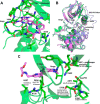
Binding of BMS-066 to Tyk2 pseudokinase domain. A, interactions of BMS-066 (carbons colored in magenta, oxygens in red, nitrogens in blue) with Tyk2 pseudokinase domain (carbons in green); B, superposition of Tyk2 pseudokinase domain/BMS-066 structure (green) with JAK1 pseudokinase domain (blue; PDB code 4L00) and JAK2 JH2 (magenta; PDB code 4FVQ) to show similar fold; C, Tyk2 pseudokinase domain structure bound with BMS-066, highlighting residues that are compared with representation in kinome. Percentages are calculated as number of kinases that possess a given residue out of 491 kinases.
FIGURE 6.
FIGURE 6.
A potential binding model of Compound 1 in the Tyk2 pseudokinase domain. Interactions of Compound 1 (carbons colored in magenta, oxygens in red, nitrogens in blue) with Tyk2 pseudokinase domain (carbons in green). Hydrogen bonds are noted by dashed lines.
FIGURE 7.
FIGURE 7.
Unique features of pseudokinase domain of Tyk2 compared with other JAK members. A, Tyk2 pseudokinase domain structure bound with BMS-066 highlighting residues that are compared with a representation in the JAK pseudokinase domain family. B, superposition of Tyk2 pseudokinase domain/BMS-066 structure (green) with JAK1 pseudokinase domain (magenta; PDB code 4L00) and JAK2 JH2 (yellow; PDB code 4FVQ), highlighting residues that may be involved in stabilization and triggering release of the autoinhibitory hold of the pseudokinase domain over the kinase domain.

References

    1. Miossec P., Kolls J.K. (2012) Targeting IL-17 and TH17 cells in chronic inflammation. Nat. Rev. Drug Discov. 11, 763–776 - PubMed
    1. Volpe E., Servant N., Zollinger R., Bogiatzi S. I., Hupé P., Barillot E., Soumelis V. (2008) A critical function for transforming growth factor-β, interleukin 23 and proinflammatory cytokines in driving and modulating human T(H)-17 responses. Nat. Immunol. 9, 650–657 - PubMed
    1. Geremia A., Arancibia-Cárcamo C. V., Fleming M. P., Rust N., Singh B., Mortensen N. J., Travis S. P., Powrie F. (2011) IL-23-responsive innate lymphoid cells are increased in inflammatory bowel disease. J. Exp. Med. 208, 1127–1133 - PMC - PubMed
    1. Sandborn W. J., Gasink C., Gao L. L., Blank M. A., Johanns J., Guzzo C., Sands B. E., Hanauer S. B., Targan S., Rutgeerts P., Ghosh S., de Villiers W. J., Panaccione R., Greenberg G., Schreiber S., Lichtiger S., Feagan B. G., and CERTIFI Study Group (2012) Ustekinumab induction and maintenance therapy in refractory Crohn's disease. N. Engl. J. Med. 367, 1519–1528 - PubMed
    1. Strober B. E., Crowley J. J., Yamauchi P. S., Olds M., Williams D. A. (2011) Efficacy and safety results from a phase III, randomized controlled trial comparing the safety and efficacy of briakinumab with etanercept and placebo in patients with moderate to severe chronic plaque psoriasis. Br. J. Dermatol. 165, 661–668 - PubMed