. 2015 Jun;89(11):5788-800.
doi: 10.1128/JVI.00406-15. Epub 2015 Mar 11.

Murine Gammaherpesvirus 68 ORF48 Is an RTA-Responsive Gene Product and Functions in both Viral Lytic Replication and Latency during In Vivo Infection

Affiliations

Murine Gammaherpesvirus 68 ORF48 Is an RTA-Responsive Gene Product and Functions in both Viral Lytic Replication and Latency during In Vivo Infection

Jing Qi et al. J Virol. 2015 Jun.

Abstract

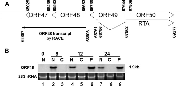
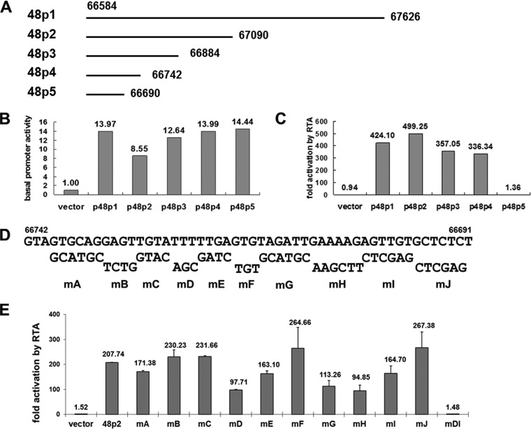
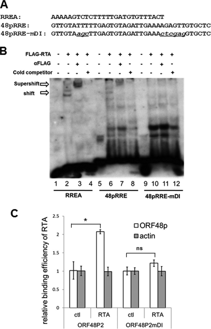
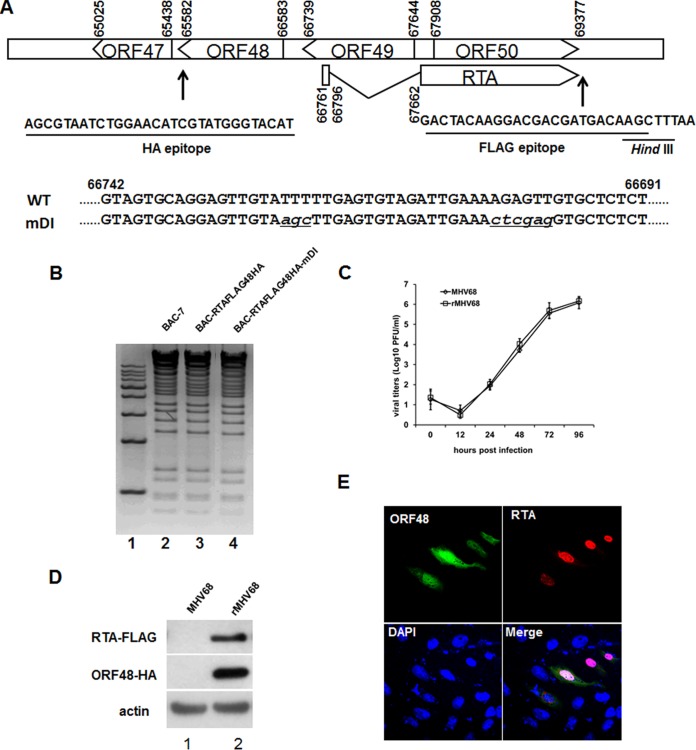
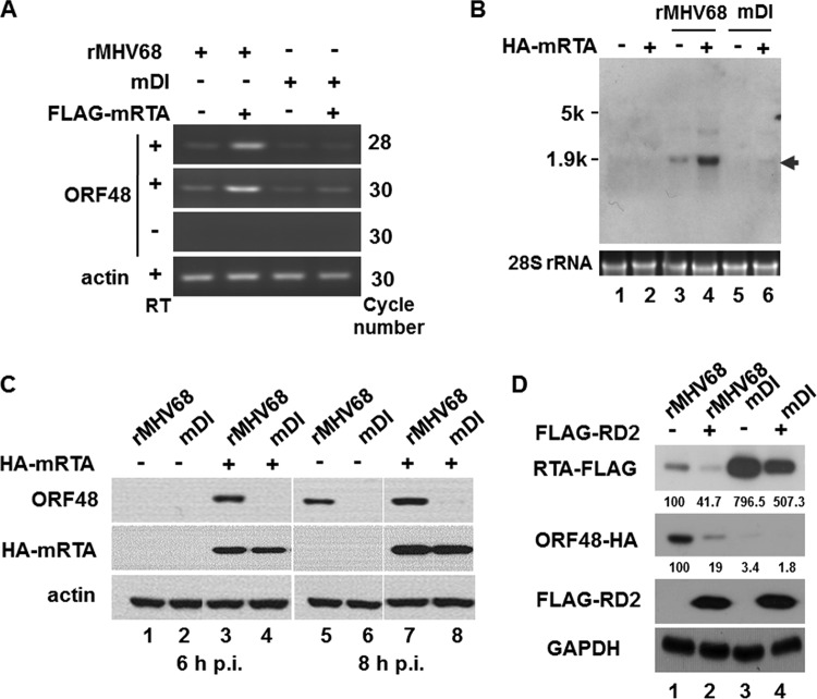
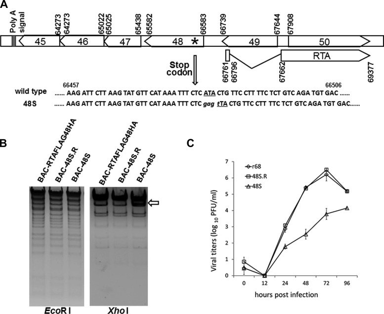
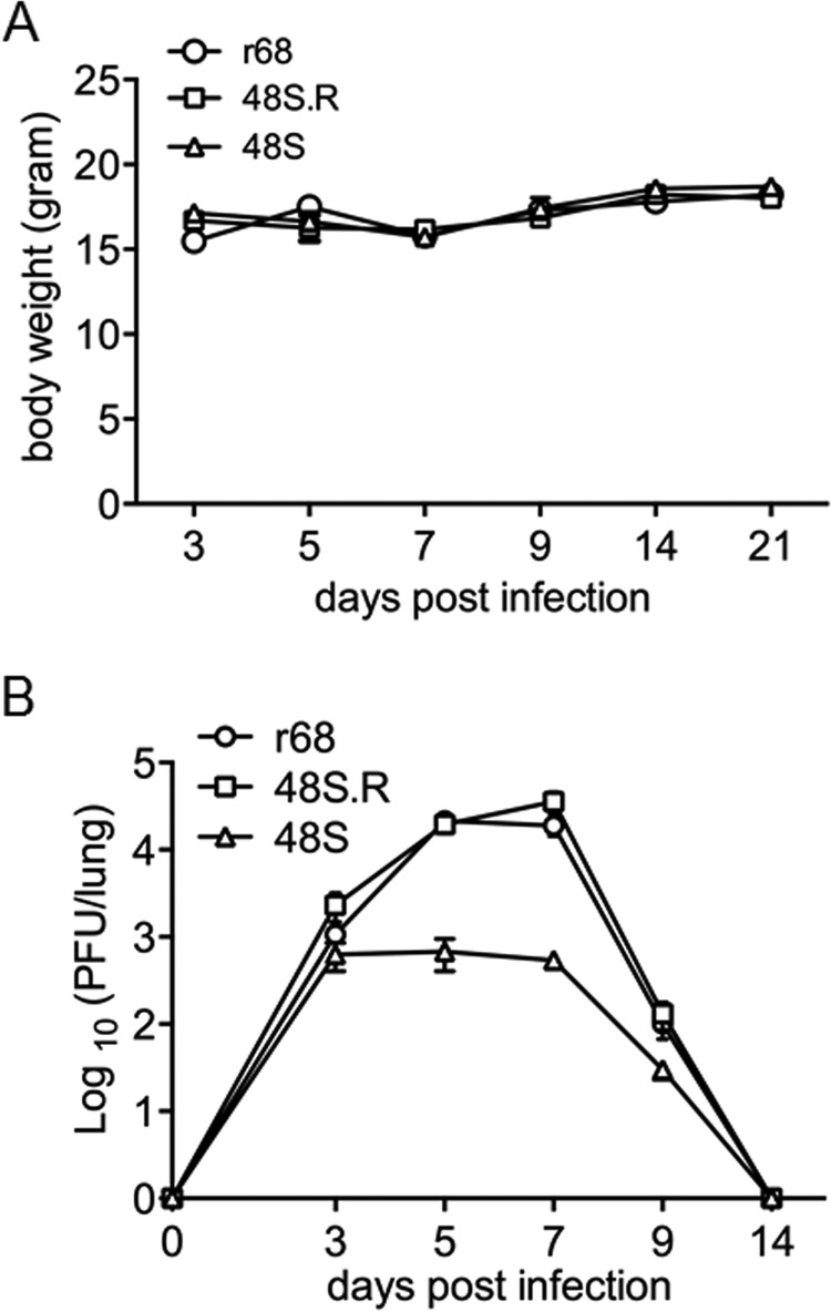
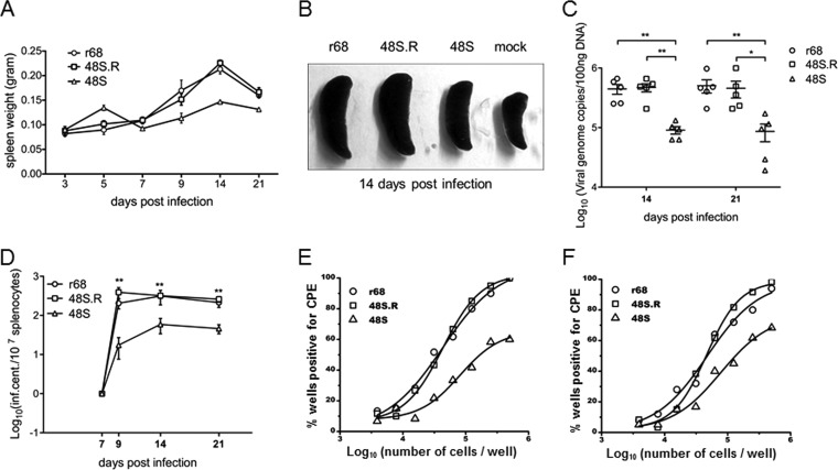
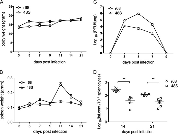
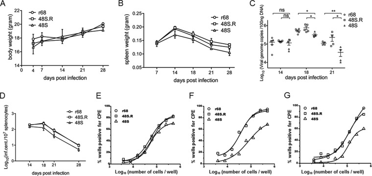
Replication and transcription activator (RTA) of gammaherpesvirus is an immediate early gene product and regulates the expression of many downstream viral lytic genes. ORF48 is also conserved among gammaherpesviruses; however, its expression regulation and function remained largely unknown. In this study, we characterized the transcription unit of ORF48 from murine gammaherpesvirus 68 (MHV-68) and analyzed its transcriptional regulation. We showed that RTA activates the ORF48 promoter via an RTA-responsive element (48pRRE). RTA binds to 48pRRE directly in vitro and also associates with ORF48 promoter in vivo. Mutagenesis of 48pRRE in the context of the viral genome demonstrated that the expression of ORF48 is activated by RTA through 48pRRE during de novo infection. Through site-specific mutagenesis, we generated an ORF48-null virus and examined the function of ORF48 in vitro and in vivo. The ORF48-null mutation remarkably reduced the viral replication efficiency in cell culture. Moreover, through intranasal or intraperitoneal infection of laboratory mice, we showed that ORF48 is important for viral lytic replication in the lung and establishment of latency in the spleen, as well as viral reactivation from latency. Collectively, our study identified ORF48 as an RTA-responsive gene and showed that ORF48 is important for MHV-68 replication both in vitro and in vivo.

Importance: The replication and transcription activator (RTA), conserved among gammaherpesviruses, serves as a molecular switch for the virus life cycle. It works as a transcriptional regulator to activate the expression of many viral lytic genes. However, only a limited number of such downstream genes have been uncovered for MHV-68. In this study, we identified ORF48 as an RTA-responsive gene of MHV-68 and mapped the cis element involved. By constructing a mutant virus that is deficient in ORF48 expression and through infection of laboratory mice, we showed that ORF48 plays important roles in different stages of viral infection in vivo. Our study provides insights into the transcriptional regulation and protein function of MHV-68, a desired model for studying gammaherpesviruses.


Figures

FIG 1
FIG 1
Characterization of the MHV-68 ORF48. (A) Schematic diagram of the locus containing RTA and ORF48 on the MHV-68 genome. Transcription initiation and termination sites of the ORF48 transcripts as mapped by 5′ and 3′ RACE assays are also shown. A total of six clones from 5′ RACE were sequenced; the transcription initiation sites were mapped to nt 66605 in four clones and to nt 66603 in two other clones. A total of six clones from 3′ RACE were sequenced; the transcription termination sites were mapped to nt 64867 in five clones and to nt 64869 in one additional clone. (B) MHV-68 ORF48 is expressed as an early gene. Cells were infected by MHV-68 at an MOI of 2 in the presence of CHX or PAA or left untreated. Total RNA was collected at 0, 8, 12, and 24 h postinfection and analyzed by Northern blotting with a probe targeting the ORF48 coding region. N, none treated; C, CHX treated; P, PAA treated.
FIG 2
FIG 2
RTA activates the ORF48 promoter in the reporter assay. (A) Diagram of the 1-kb ORF48 promoter and a series of 5′ deletion fragments. (B and C) Dual-luciferase reporter assays showing the basal activities of the ORF48 promoter constructs and their activation by RTA. (D) Diagram of systematic site-directed mutations of 48pRRE. The wild-type sequences are shown on the top, and the mutated sequences and the names of the corresponding reporter plasmids (mA to mJ) are shown below. (E) Reporter assays with the 48pRRE mutants identified nucleotides critical for 48pRRE function. The fold activation by RTA for the vector is significantly different from that for each of the ORF48 promoter reporter plasmids except mDI (P < 0.01). The difference between activation for 48p2 and that for each of mD, mE, mG, mH, and mI is also significant (P < 0.01).
FIG 3
FIG 3
RTA binds to 48pRRE in vitro and in vivo. (A) Sequences of biotin-labeled probes used in EMSA. (B) RTA specifically binds to 48pRRE in EMSA. Nuclear extract from pFLAGCMV2-RTA-transfected 293T cells was incubated with biotin-labeled probes (RREA, 48pRRE, and 48pRRE-mDI). In supershift reactions, anti-FLAG antibody was added; in competition reactions, a 200-fold excess amount of unlabeled RREA oligonucleotides was added. (C) RTA associates with ORF48 promoter in a ChIP assay. 293T cells were transfected with ORF48 promoter plasmids (p48p2 or p48p2-mDI) and pFLAGCMV2-RTA (or empty vector pFLAGCMV2 as a control). A ChIP assay was performed by cross-linking DNA-protein complexes and immunoprecipitation with anti-FLAG antibody-conjugated beads. Quantitative PCR was performed with primers specific for the ORF48 promoter region or actin coding region as a control. Three independent experiments were performed, and the relative amount of immunoprecipitated DNA was first normalized to the input DNA and then compared to that of control vector (pFLAGCMV2)-transfected cells. *, P < 0.05; ns, P > 0.05.
FIG 4
FIG 4
Construction of an RTA and ORF48 doubly tagged MHV-68 virus and an mDI mutant virus. (A) Diagram of the sequences and locations of the FLAG and HA tags on the viral genome, as well as the sequence of mDI mutant virus in comparison to that of the wild-type virus. (B) Restriction enzyme digestion analysis of wild-type MHV-68 BAC-7, BAC-RTAFLAG-48HA, and BAC-RTAFLAG-48HA-mDI. (C) Multiple-step growth curves of wild-type MHV-68 and doubly tagged virus (rMHV68). (D) Detection of FLAG-tagged RTA and HA-tagged ORF48 during wild-type MHV-68 and rMHV68 de novo infection. Cells were infected at an MOI of 3 for 12 h and then collected for Western blotting. (E) Cellular localization of ORF48 and RTA during de novo infection. Vero cells were infected with rMHV68 for 12 h and then subjected to an indirect immunofluorescence assay. Green, ORF48; red, RTA.
FIG 5
FIG 5
RTA activates transcription and expression of ORF48 via 48pRRE from MHV-68 genome. (A and B) Recombinant virus rMHV68 or rMHV68-mDI was used to infect 293T cells which had been transfected with pCMVHA-mRTA (or pCMV-HA) 24 h before infection. Four hours after infection, total RNA was isolated for analyzing ORF48 expression by RT-PCR with primers specific to the ORF48 coding region (nt 65852 to 65997) (A) or Northern blotting with a probe targeting the ORF48 coding region (B). (C) RTA activated the expression of endogenous ORF48 protein via 48pRRE. Transfection and infection were performed as described above, and cells were harvested at the indicated time points, followed by Western blotting. (D) A dominant negative form of RTA (FLAG-RD2) inhibited the expression of endogenous ORF48. Expression plasmid FLAG-RD2 or a vector control was transfected into cells 24 h prior to infection with rMHV68 or rMHV68-mDI. Cells were collected at 8 h after infection and analyzed by Western blotting with antibodies against the HA epitope (ORF48) and FLAG epitope (endogenous RTA and FLAG-RD2). The relative expression levels of endogenous RTA-FLAG and ORF48-HA were calculated with ImageJ.
FIG 6
FIG 6
Construction and characterization of an MHV-68 ORF48-null virus. (A) Schematic illustration of the MHV-68 locus harboring the mutation in the ORF48-null mutant. Briefly, nucleotide A at position 66487 is mutated to T in order to form a translation termination codon on the viral genome (102 nt downstream of the translation start codon for ORF48). (B) Restriction enzyme digestion analysis of BAC-RTAFLAG-48HA and the ORF48-null and revertant mutant. (C) Multiple-step growth curves of the recombinant viruses. NIH 3T3 cells were infected at an MOI of 0.01, cells and supernatants were harvested at the indicated time points postinfection, and viral titers were determined by plaque assay.
FIG 7
FIG 7
MHV-68 ORF48-null virus displayed a significant growth defect in the lung of infected mice. Results from one representative experiment are shown. (A) Body weights of BALB/c mice infected with 500 PFU of each indicated virus. (B) Infectious virus load in the lungs. The lungs were removed from infected mice at the indicated time points postinfection, and tissue homogenates were used to measure viral titers by standard plaque assays. Each time point represents data from five animals, and error bars represent standard deviations.
FIG 8
FIG 8
ORF48 is important for MHV-68 latent infection in the spleen. Results from one representative experiment are shown. (A) The spleen weights of mice infected with 500 PFU of viruses at the indicated days postinfection. (B) Representative images of spleens from mice infected at 14 days postinfection. (C) Quantitative analysis of the viral DNA load in the splenocytes from r68-, 48S.R-, or 48S-infected mice. Real-time PCR was performed using primers specific for MHV-68 ORF65. All time points represent results from five animals, and error bars represent standard deviations. (D to F) Levels of reactivating viruses in the spleens of infected mice. Single-cell suspensions were obtained from spleens harvested at the indicated days postinfection and subjected to infectious center assays to determine the virus reactivation efficiency (D). Ex vivo limiting dilution assays were also performed with the same single-cell suspensions harvested at day 14 (E) and day 21 (F) postinfection. For each virus infection at each time point, splenocytes from five infected mice (5 × 10⁶ cells each) were pooled and analyzed in the limiting dilution assay. *, P < 0.05; **, P < 0.01 (comparison between results for r68 or 48S.R and 48S).
FIG 9
FIG 9
A higher dose of inoculum intranasally does not rescue the impairment displayed by ORF48-null mutation in either lytic replication or latency. (A) Body weights of BALB/c mice infected with 5 × 10⁴ PFU of each indicated virus. (B) Viral titers in lungs from infected mice were determined at 3, 5, 7, and 9 days p.i. (C) Measurement of spleen weights at indicated days after inoculation. (D) Levels of reactivating viruses in the spleens of infected mice. Single-cell suspensions were obtained from spleens harvested at days 14 and 21 postinfection and subjected to infectious center assays. For all experiments, each time point represents data from five animals, and error bars represent standard deviations. *, P < 0.05; **, P < 0.01 (comparison between results for r68 and 48S).
FIG 10
FIG 10
ORF48 is important for MHV-68 latent infection during intraperitoneal inoculation. Results from one representative experiment are shown. (A) Body weights of mice at indicated days after infection with 500 PFU viruses. (B) Spleen weights at indicated days postinfection. (C) Quantitation of viral DNA in the splenocytes. ns, not significant (P > 0.05; *, P < 0.05; **, P < 0.01, comparing the results between r68 or 48S.R and 48S. (D to G) Levels of viral reactivation from splenocytes. Single-cell suspensions were obtained from the spleens harvested at days 14, 18, 21, and 28 postinfection and examined for reactivation efficiency by infectious center assays (D). Single-cell suspensions from day 18 (E), day 21(F), and day 28 (G) were also subjected to ex vivo limiting dilution assays. For each virus infection at each time point, splenocytes from five infected mice (5 × 10⁶ cells each) were pooled and analyzed.

Similar articles

Cited by

References

    1. Stevenson PG. 2004. Immune evasion by gamma-herpesviruses. Curr Opin Immunol 16:456–462. doi:10.1016/j.coi.2004.05.002. - DOI - PubMed
    1. Jenner RG, Albà MM, Boshoff C, Kellam P. 2001. Kaposi's sarcoma-associated herpesvirus latent and lytic gene expression as revealed by DNA arrays. J Virol 75:891–902. doi:10.1128/JVI.75.2.891-902.2001. - DOI - PMC - PubMed
    1. Samols MA, Hu J, Skalsky RL, Renne R. 2005. Cloning and identification of a microRNA cluster within the latency-associated region of Kaposi's sarcoma-associated herpesvirus. J Virol 79:9301–9305. doi:10.1128/JVI.79.14.9301-9305.2005. - DOI - PMC - PubMed
    1. Dittmer D, Lagunoff M, Renne R, Staskus K, Haase A, Ganem D. 1998. A cluster of latently expressed genes in Kaposi's sarcoma-associated herpesvirus. J Virol 72:8309–8315. - PMC - PubMed
    1. Bais C, Santomasso B, Coso O, Arvanitakis L, Raaka EG, Gutkind JS, Asch AS, Cesarman E, Gerhengorn MC, Mesri EA. 1998. G-protein-coupled receptor of Kaposi's sarcoma-associated herpesvirus is a viral oncogene and angiogenesis activator. Nature 391:86–89. doi:10.1038/34193. - DOI - PubMed

Publication types

MeSH terms

LinkOut - more resources