. 2015 Apr 15;194(8):3687-96.
doi: 10.4049/jimmunol.1401803. Epub 2015 Mar 11.

Coexpression of TIGIT and FCRL3 identifies Helios+ human memory regulatory T cells

Affiliations

Coexpression of TIGIT and FCRL3 identifies Helios+ human memory regulatory T cells

Khalid Bin Dhuban et al. J Immunol. .

Abstract

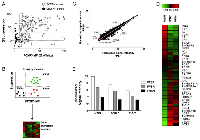
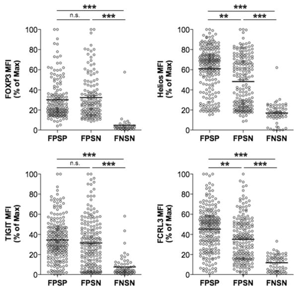
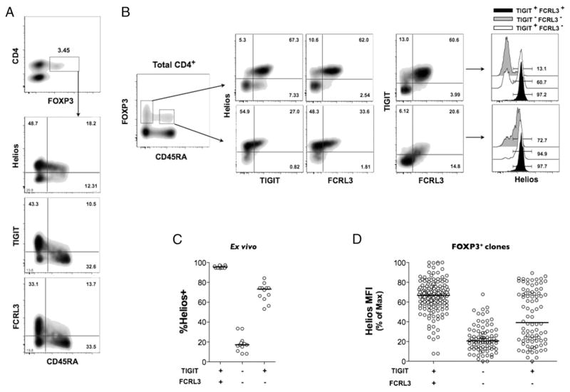
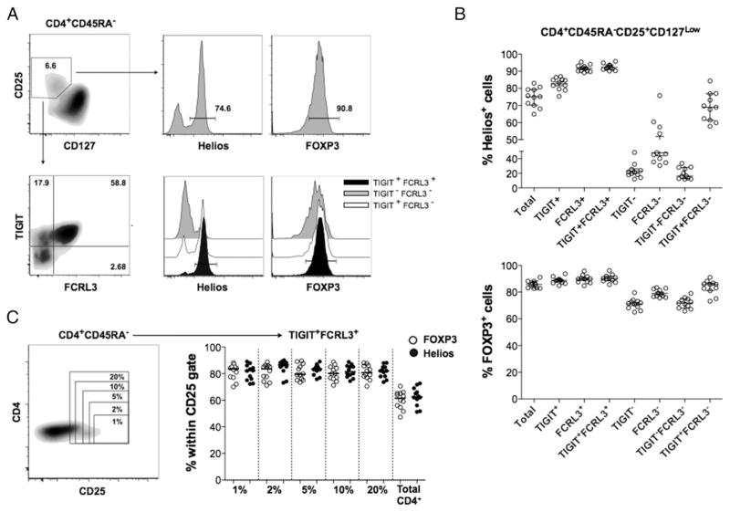
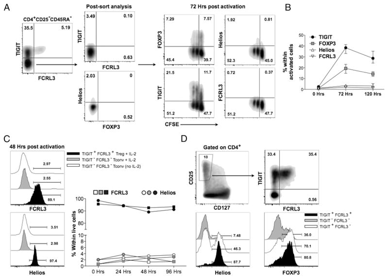
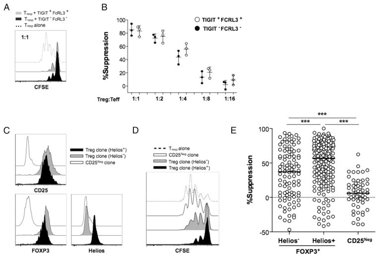
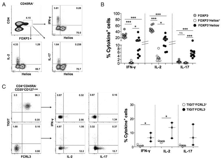
Two distinct subsets of CD4(+)Foxp3(+) regulatory T (Treg) cells have been described based on the differential expression of Helios, a transcription factor of the Ikaros family. Efforts to understand the origin and biological roles of these Treg populations in regulating immune responses have, however, been hindered by the lack of reliable surface markers to distinguish and isolate them for subsequent functional studies. Using a single-cell cloning strategy coupled with microarray analysis of different Treg functional subsets in humans, we identify the mRNA and protein expression of TIGIT and FCRL3 as a novel surface marker combination that distinguishes Helios(+)FOXP3(+) from Helios(-)FOXP3(+) memory cells. Unlike conventional markers that are modulated on conventional T cells upon activation, we show that the TIGIT/FCRL3 combination allows reliable identification of Helios(+) Treg cells even in highly activated conditions in vitro as well as in PBMCs of autoimmune patients. We also demonstrate that the Helios(-)FOXP3(+) Treg subpopulation harbors a larger proportion of nonsuppressive clones compared with the Helios(+)FOXP3(+) cell subset, which is highly enriched for suppressive clones. Moreover, we find that Helios(-) cells are exclusively responsible for the productions of the inflammatory cytokines IFN-γ, IL-2, and IL-17 in FOXP3(+) cells ex vivo, highlighting important functional differences between Helios(+) and Helios(-) Treg cells. Thus, we identify novel surface markers for the consistent identification and isolation of Helios(+) and Helios(-) memory Treg cells in health and disease, and we further reveal functional differences between these two populations. These new markers should facilitate further elucidation of the functional roles of Helios-based Treg heterogeneity.


Conflict of interest statement

Disclosures

The authors have no financial conflicts of interest.

Figures

FIGURE 1
FIGURE 1
Gene expression analysis of suppressive versus nonsuppressive FOXP3+ clones. Illumina BeadChip analysis was used for total mRNA prepared from individual clones representing three populations: 1) suppressive and 2) nonsuppressive FOXP3+ clones generated from CD4+CD25high cells, and 3) control FOXP3 clones generated from CD4+CD25 cells. (A) Correlation of suppressive potency with FOXP3 expression levels in primary FOXP3+ and FOXP3 (CD25) clones generated from three healthy donors. The dotted lines represent the cutoffs for FOXP3 mean fluorescence intensity (MFI) (x-axis) and percentage suppression (y-axis) determined through the overall FOXP3 MFI and suppressive capacity of the control FOXP3 clones generated from CD4+CD25 cells. (B) Schematic illustrating the process of selection of representative clones. (C) Scatter plot showing normalized gene expression level in suppressive versus nonsuppressive clones in the resting state. (D) Heat map comparing variations in the expression of selected Treg- and Tconv-associated genes relative to the median mRNA levels across the three subsets. (E) Relative mRNA expression levels of TIGIT, FCRL3, and Helios in the three functional categories. Representative clones were derived from two healthy donors, and two to three similar clones/subset/donor were pooled to obtain sufficient mRNA.
FIGURE 2
FIGURE 2
FPSN clones comprise majorly Helios clones and display a reduced FCRL3 expression. Primary Treg and Tconv clones were generated by single cell cloning of FACS-sorted CD25high and CD25 cells of four healthy individuals. Marker expression analysis was performed immediately after the harvest on days 22–24, and 4-d CFSE-based suppression assays were carried out using allogeneic CD4+CD25 cells as responders at a 1:1 Treg/Tresp ratio in the presence of irradiated PBMCs and anti-CD3 (30 ng/ml). Suppression was measured relative to the division index of the unsuppressed Tresp-alone control. Shown are the expression levels of FOXP3, Helios, TIGIT, and FCRL3 in FPSP, FPSN, and FNSN clones immediately after harvest. Statistical analysis was done with the one-way ANOVA followed by a Tukey posttest. **p ≤ 0.01, ***p ≤ 0.001. n.s., not significant.
FIGURE 3
FIGURE 3
The combined expression of TIGIT and FCRL3 discriminates between Helios+ and Helios subsets in memory FOXP3+ cells. (AC) PBMCs from healthy subjects were analyzed ex vivo by flow cytometry. (A) Expression of TIGIT, FCRL3, and Helios on naive and memory CD4+FOXP3+ populations. (B) Representative plots showing the correlation of TIGIT and FCRL3 expression with Helios expression in naive versus memory CD4+ FOXP3+ cells. (C) Combined analysis of 11 healthy individuals showing the application of TIGIT and FCRL3 in the identification Helios+ and Helios subsets within memory FOXP3+ cells. (D) Applicability of TIGIT/FCRL3 combination in discriminating Helios subsets in FOXP3+ clones generated from seven healthy donors (n = 299 clones).
FIGURE 4
FIGURE 4
TIGIT/FCRL3 combination provides a reliable surface marker for the isolation of Helios+ and Helios memory Treg cells. PBMCs from 11 healthy subjects were analyzed ex vivo by flow cytometry. (A) Representative FACS plots showing the application of the TIGIT/FCRL3 marker combination in distinguishing Helios subsets within memory CD4+CD25+CD127low cells. (B) Identification of Helios subsets by TIGIT/FCRL3 surface markers is precisely reproducible in healthy samples with a wide range of Helios expression. Shown is the frequency of Helios+ (top) and FOXP3+ (bottom) cells in Treg populations gated using the conventional markers (CD25+CD127low; referred to as Total) compared with further gating using different combinations of TIGIT and FCRL3. (C) The TIGIT/FCRL3 combination allows the identification of consistently enriched FOXP3+Helios+ populations with less stringent gating on CD25. Shown is the frequency of FOXP3+ and Helios+ cells in TIGIT+FCRL3+ cells obtained from variably stringent CD25+ gates on memory CD4+ cells.
FIGURE 5
FIGURE 5
The TIGIT/FCRL3 marker expression can reliably identify Helios+ and Helios Treg cell subsets in inflammatory contexts. (A) FACS-sorted TIGITFCRL3 memory CD4+CD25 cells from PBMCs of a healthy donor were labeled with CFSE and activated in vitro with anti-CD3/anti-CD28–coated beads at a ratio of two beads/one cell for 5 d. Shown are representative FACS plots and (B) the kinetics of activation-induced marker upregulation in three separate experiments on three healthy individuals. (C) Total CD4+CD25TIGITFCRL3 cells from PBMCs activated in vitro with anti-CD3/anti-CD28–coated beads at a ratio of two beads/one cell with or without recombinant human IL-2 in the presence of irradiated autologous feeders for 4 d. Shown is the expression of FCRL3 and Helios on the activated CD4+CD25TIGITFCRL3 cells compared with FACS-sorted CD4+CD25+CD127lowTIGIT+FCRL3+ cells plated in parallel. (D) Whole PBMCs from a healthy individual were stimulated with anti-CD3/anti-CD28–coated beads at a ratio of two beads/one cell for 72 h. Representative FACS plots from similar experiments on three healthy donors are shown.
FIGURE 6
FIGURE 6
An increased frequency of nonsuppressive clones is found within the FOXP3+Helios subset. The suppressive potency of Helios+ and Helios Treg cells was compared ex vivo (A and B) and in primary clones (CE). For ex vivo suppression assays, CD4+CD25+CD127lowTIGIT+FCRL3+ and CD4+CD25+CD127lowTIGITFCRL3 cells were FACS sorted and tested for the capacity to suppress the proliferation of CFSE-labeled CD4+CD25 Tresp cells stimulated with soluble anti-CD3 and irradiated PBMCs for 4 d. (A) Representative CFSE dilution histograms showing suppression of Tresp cells. (B) Combined suppression analysis from three different experiments using cells from three different healthy individuals. (CE) Primary Treg and Tconv clones were generated by single-cell cloning of FACS-sorted CD25high and CD25 cells of seven healthy individuals. Marker expression was performed immediately after the harvest on days 22–24, and 4-d CFSE-based suppression assays were carried out using allogeneic CD4+CD25 cells as responders at a 1:1 Treg/Tresp ratio in the presence of irradiated PBMCs and anti-CD3 (30 ng/ml). (C) The expression of CD25, FOXP3, and Helios in representative clones at harvest. (D and E) Suppressive potency of FOXP3+Helios+ (n = 196 clones), FOXP3+Helios (n = 103 clones), and FOXP3 (n = 59 clones). Suppression was measured relative to the division index of the unsuppressed Tresp-alone control. Statistical analysis was done with the one-way ANOVA followed by a Tukey posttest. ***p ≤ 0.001.
FIGURE 7
FIGURE 7
Inflammatory cytokine production in FOXP3+ cells is restricted to the Helios fraction. (A) PBMCs were incubated ex vivo with PMA (25 ng/ml), ionomycin (1 μg/ml), and GolgiStop for 4 h, followed by intracellular cytokine staining. Shown are representative flow cytometry plots (A) and the frequency of cytokine-producing cells in the indicated subsets (B) analyzed from 11 different healthy samples. (C) Cytokine production in healthy FACS-sorted TIGIT+FCRL3+ versus TIGITFCRL3 Treg cells (CD4+CD45RACD25+CD127low). Statistical analysis was done with the one-way ANOVA followed by a Tukey posttest (B) or with the Student t test (C). *p ≤ 0.05, ***p ≤ 0.001. n.s., not significant.

References

    1. Sakaguchi S, Miyara M, Costantino CM, Hafler DA. FOXP3+ regulatory T cells in the human immune system. Nat Rev Immunol. 2010;10:490–500. - PubMed
    1. d’Hennezel E, Bin Dhuban K, Torgerson T, Piccirillo CA. The immunogenetics of immune dysregulation, polyendocrinopathy, enteropathy, X linked (IPEX) syndrome. J Med Genet. 2012;49:291–302. - PubMed
    1. Buckner JH. Mechanisms of impaired regulation by CD4+CD25+ FOXP3+ regulatory T cells in human autoimmune diseases. Nat Rev Immunol. 2010;10:849–859. - PMC - PubMed
    1. Liu W, Putnam AL, Xu-Yu Z, Szot GL, Lee MR, Zhu S, Gottlieb PA, Kapranov P, Gingeras TR, Fazekas de St Groth B, et al. CD127 expression inversely correlates with FoxP3 and suppressive function of human CD4+ T reg cells. J Exp Med. 2006;203:1701–1711. - PMC - PubMed
    1. Gavin MA, Torgerson TR, Houston E, DeRoos P, Ho WY, Stray-Pedersen A, Ocheltree EL, Greenberg PD, Ochs HD, Rudensky AY. Single-cell analysis of normal and FOXP3-mutant human T cells: FOXP3 expression without regulatory T cell development. Proc Natl Acad Sci USA. 2006;103:6659–6664. - PMC - PubMed

Publication types

MeSH terms

Associated data