Review
. 2015 Feb 13:9:10.
doi: 10.3389/fnana.2015.00010. eCollection 2015.

GDNF-based therapies, GDNF-producing interneurons, and trophic support of the dopaminergic nigrostriatal pathway. Implications for Parkinson's disease

Affiliations
Review

GDNF-based therapies, GDNF-producing interneurons, and trophic support of the dopaminergic nigrostriatal pathway. Implications for Parkinson's disease

Xavier d'Anglemont de Tassigny et al. Front Neuroanat. .

Abstract

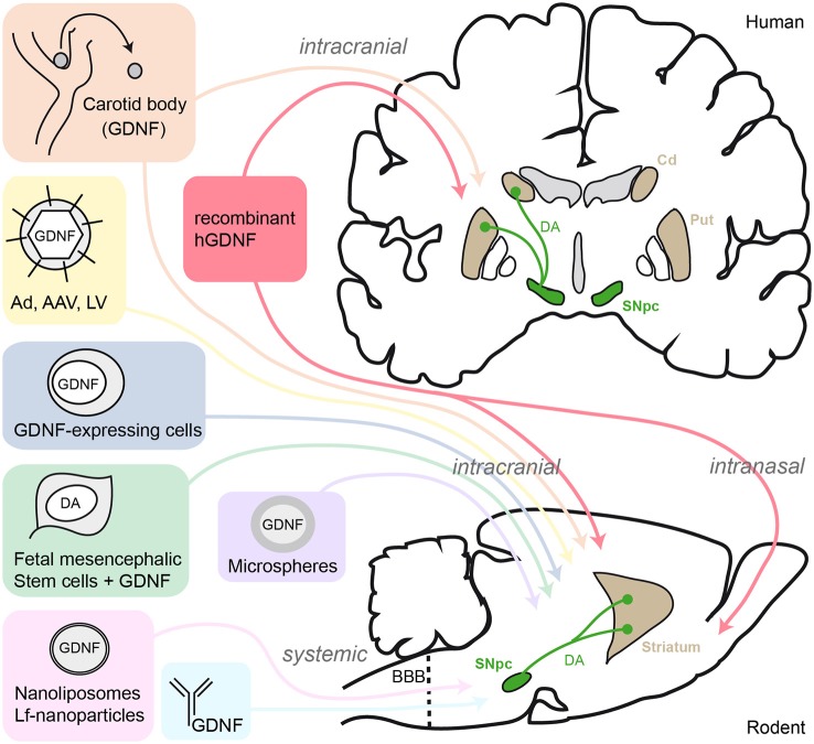
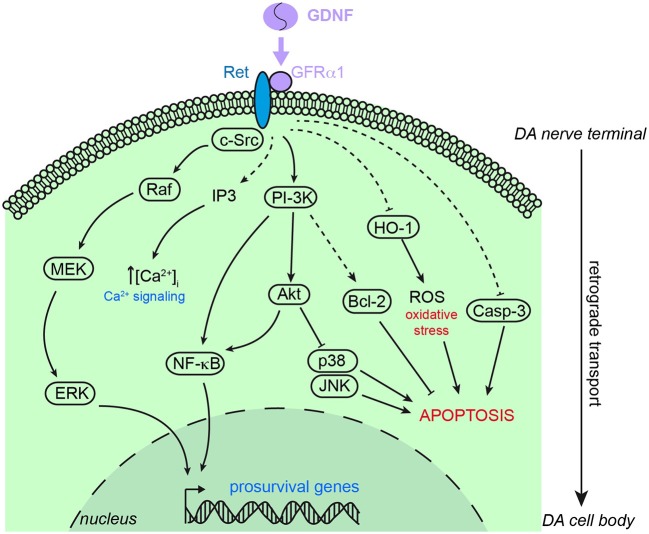
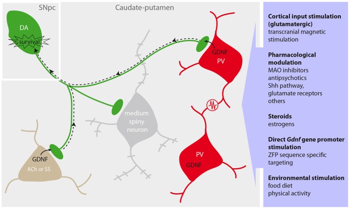
The glial cell line-derived neurotrophic factor (GDNF) is a well-established trophic agent for dopaminergic (DA) neurons in vitro and in vivo. GDNF is necessary for maintenance of neuronal morphological and neurochemical phenotype and protects DA neurons from toxic damage. Numerous studies on animal models of Parkinson's disease (PD) have reported beneficial effects of GDNF on nigrostriatal DA neuron survival. However, translation of these observations to the clinical setting has been hampered so far by side effects associated with the chronic continuous intra-striatal infusion of recombinant GDNF. In addition, double blind and placebo-controlled clinical trials have not reported any clinically relevant effect of GDNF on PD patients. In the past few years, experiments with conditional Gdnf knockout mice have suggested that GDNF is necessary for maintenance of DA neurons in adulthood. In parallel, new methodologies for exogenous GDNF delivery have been developed. Recently, it has been shown that a small population of scattered, electrically interconnected, parvalbumin positive (PV+) GABAergic interneurons is responsible for most of the GDNF produced in the rodent striatum. In addition, cholinergic striatal interneurons appear to be also involved in the modulation of striatal GDNF. In this review, we summarize current knowledge on brain GDNF delivery, homeostasis, and its effects on nigrostriatal DA neurons. Special attention is paid to the therapeutic potential of endogenous GDNF stimulation in PD.

Keywords: GDNF; Parkinson disease; dopaminergic system; mouse models; neurotrophic factors; nigrostriatal pathway; parvalbumin interneurons; striatum.


Figures

Figure 1
Figure 1
Schematic summary of GDNF-delivery strategies tested in human PD patients and rodent models. AAV, adeno-associated virus; Ad, adenovirus; LV, lentivirus; Lf, lactoferrin; SNpc, substantia nigra pars compacta; DA, dopamine; Cd, caudate nucleus; Put, putamen; BBB, blood-brain-barrier.
Figure 2
Figure 2
Schematic representation of the main signaling pathways involved in the neuroprotective action of GDNF on dopaminergic neurons. GDNF principally stimulates the binding of GFRα1 and Ret to trigger intracellular signaling cascades leading to pro-survival genes expression, calcium signaling and pro-apoptosis factors inhibition. Akt, protein kinase B; Bcl-2, B cell lymphoma 2; Casp-3, caspase 3; c-Src, proto-oncogene tyrosine-protein kinase Src; ERK, extracellular signal-regulated kinase; HO1, heme oxygenase 1; IP3, inositol tris-phosphate; JNK, c-Jun N-terminal kinase; MEK, mitogen extracellular signal-regulated kinase; NF-κB, nuclear factor kappa B; PI-3K, phosphatidylinositol 3 kinase; Raf, Raf kinase; ROS, reactive oxygen species. Dashed arrows indicate indirect stimulation or inhibition.
Figure 3
Figure 3
Protection of the dopaminergic nigrostriatal pathway by striatal GDNF and activation of endogenous GDNF production. Dopaminergic (DA) neurons (green) located in the substantia nigra pars compacta (SNpc) innervate the caudate-putamen to modulate the activity of GABAergic medium spiny neurons (gray), parvalbumin (PV)-positive interneurons (red) and other cholinergic (ACh) or somatostatin (SS) interneurons (brown). PV neurons form an ensemble of synchronized cells through multiple dendrodendritic electrical synapses (resistance in the scheme), and release GDNF at the nerve terminals to provide trophic support to DA neurons via retrograde signaling (dotted arrow). Proposed strategies to enhance the endogenous GDNF production are summarized in the purple box (right).

References

    1. Airaksinen M. S., Saarma M. (2002). The GDNF family: signalling, biological functions and therapeutic value. Nat. Rev. Neurosci. 3, 383–394. 10.1038/nrn812 - DOI - PubMed
    1. Åkerud P., Canals J. M., Snyder E. Y., Arenas E. (2001). Neuroprotection through delivery of glial cell line-derived neurotrophic factor by neural stem cells in a mouse model of Parkinson’s disease. J. Neurosci. 21, 8108–8118. - PMC - PubMed
    1. Anastasia A., de Erausquin G. A., Wojnacki J., Mascó D. H. (2007). Protection of dopaminergic neurons by electroconvulsive shock in an animal model of Parkinson’s disease. J. Neurochem. 103, 1542–1552. 10.1111/j.1471-4159.2007.04856.x - DOI - PubMed
    1. Anastasía A., Wojnacki J., de Erausquin G. A., Mascó D. H. (2011). Glial cell-line derived neurotrophic factor is essential for electroconvulsive shock-induced neuroprotection in an animal model of Parkinson’s disease. Neuroscience 195, 100–111. 10.1016/j.neuroscience.2011.08.019 - DOI - PubMed
    1. Bar-Am O., Weinreb O., Amit T., Youdim M. B. H. (2005). Regulation of Bcl-2 family proteins, neurotrophic factors and APP processing in the neurorescue activity of propargylamine. FASEB J. 19, 1899–1901. 10.1096/fj.05-3794fje - DOI - PubMed