. 2015 Feb 16:9:11.
doi: 10.3389/fnana.2015.00011. eCollection 2015.

Ontogenesis of NADPH-diaphorase positive neurons in guinea pig neocortex

Affiliations

Ontogenesis of NADPH-diaphorase positive neurons in guinea pig neocortex

Chao Liu et al. Front Neuroanat. .

Abstract

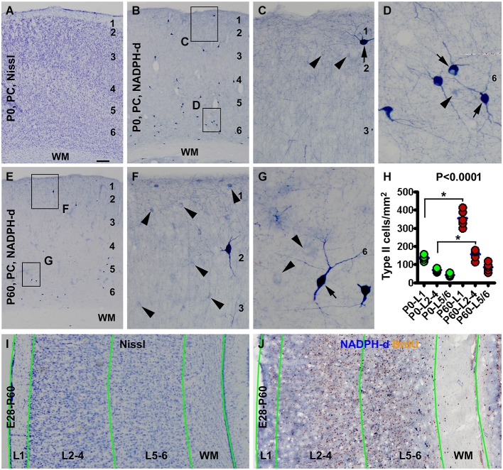
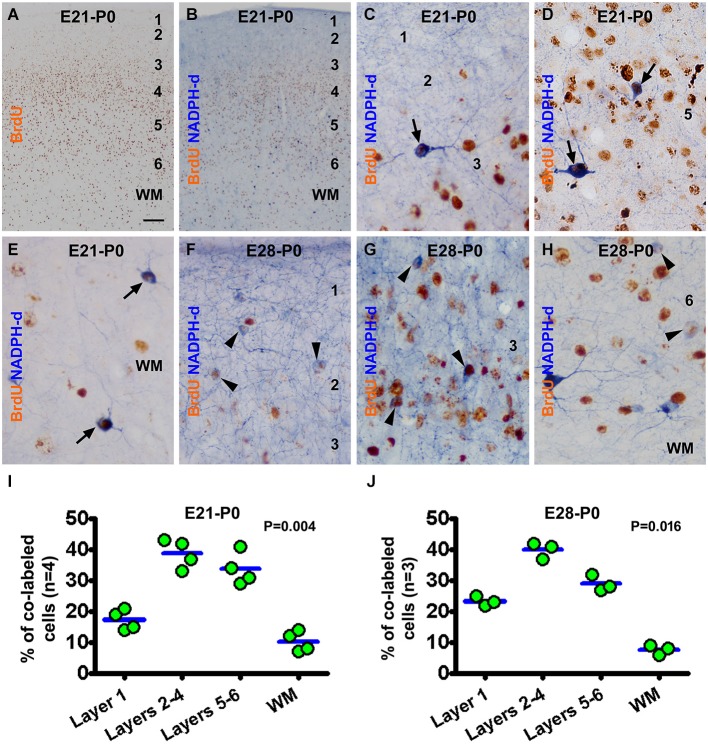
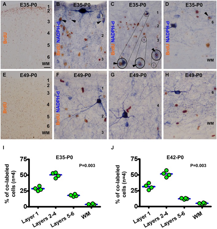
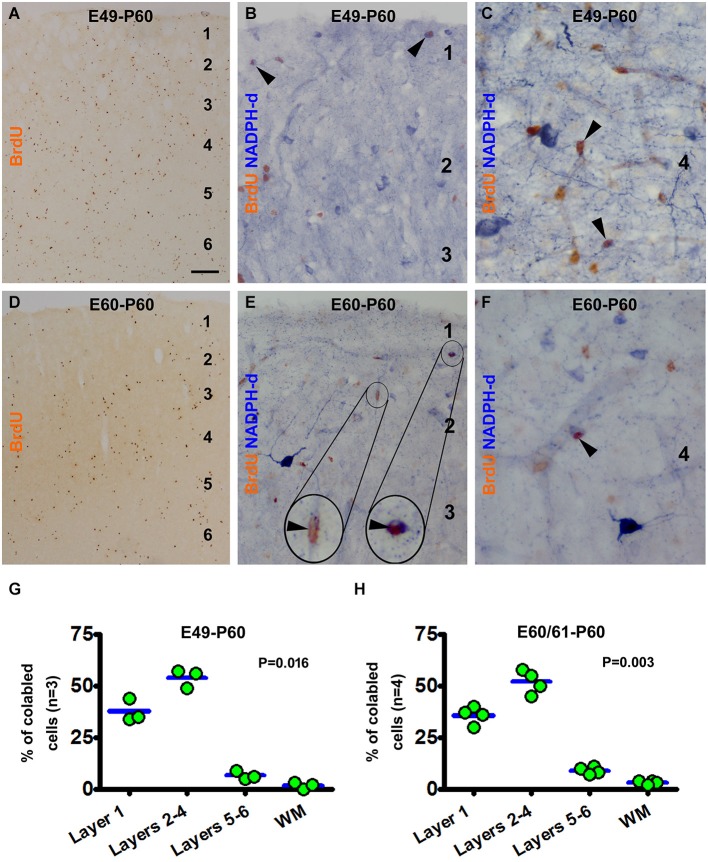
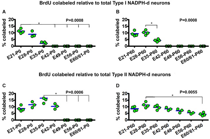
In mammalian cerebrum there exist two distinct types of interneurons expressing nitric oxide synthase (NOS). Type I neurons are large in size and exhibit heavy nicotinamide adenine dinucleotide phosphate diaphorase (NADPH-d) histochemical reaction, while type II cells are small with light NADPH-d reactivity. The time of origin of these cortical neurons relative to corticogenesis remains largely unclear among mammals. Here we explored this issue in guinea pigs using cell birth-dating and double-labeling methods. Bromodeoxyuridine (BrdU) pulse-chasing (2 doses at 50 mg/kg, 12 h apart) was given to time-pregnant mothers, followed by quantification of NADPH-d/BrdU colocalization in the parietal and temporal neocortex in offspring at postnatal day 0 (P0), P30 and P60. Type I neurons were partially colabeled with BrdU at P0, P30 and P60 following pulse-chasing at embryonic day 21 (E21), E28 and E35, varied from 2-11.3% of total population of these neurons for the three time groups. Type II neurons were partially colabeled for BrdU following pulse-chasing at E21, E28, E35 and E42 at P0 (8.6%-16.5% of total population for individual time groups). At P60, type II neurons were found to co-express BrdU (4.8-11.3% of total population for individual time groups) following pulse-chasing at E21, E28, E35, E42, E49, E56 and E60/61. These results indicate that in guinea pigs type I neurons are generated during early corticogenesis, whereas type II cells are produced over a wide prenatal time window persisting until birth. The data also suggest that type II nitrinergic neurons may undergo a period of development/differentiation, for over 1 month, before being NADPH-d reactive.

Keywords: GABAergic; corticogenesis; interneuron; neuronal development; nitric oxide.


Figures

Figure 1
Figure 1
Laminar architecture, nicotinamide adenine dinucleotide phosphate-diaphorase (NADPH-d) positive neurons in the neocortex of neonatal and 2 month-old (postnatal day 60, P60) guinea pigs, and methodological illustration for lamina-based cell count. Panel (A) shows a Nissl stain image of the parietal neocortex (PC) around the border of the primary motor (left portion) and somatosensory (right portion) cortex from a newborn (P0). Layers 1 to 6 are identifiable above the white matter (WM). Panel (B) is a low magnification image of NADPH-d neurons in the somatosensory cortex of a newborn, with the framed areas enlarged as (C) and (D). Panel (E) is a low power view of NADPH-d reactivity in the somatosensory cortex in a P60 animal, with the framed areas enlarged as (F) and (G). Examples of type I and type II NADPH-d neurons are pointed by arrows and arrowheads, respectively. The densities of type II NADPH-d neurons in layer 1 (L1), layers 2–4 (L2-4) and layers 5 and 6 (L5/6) in the parietotemporal neocortex are increased in the P60 (n = 5) relative to P0 (n = 5) groups (calculated based on the cell count data obtained from NADPH-d/BrdU dual staining preparations) (H). For counting NADPH-d positive and NADPH-d/BrdU colabeled neurons, the sampled parietal and temporal cortical regions are divided into L1, L2-4, L5/6 and white matter (WM). Borders of the laminar sectors are created in the montaged Nissl stain image of an adjacent section (I), which were copied as a template and then pasted on the montaged dual staining image (J). The size of the template is adjusted proportionally by aligning the pial and ventricular lines as closely as possible. NADPH-d neurons in each laminar sector are classified and counted on screen at high resolution. Scale bar = 100 μm in (A) applying to (B,E), equivalent to 50 μm for (I,J), 20 μm for (C,F) and 10 μm for (D,G).
Figure 2
Figure 2
Representative images and quantitative laminar analyses showing bromodeoxyuridine (BrdU) labeling and colocalization in NADPH-d neurons in neonatal guinea pig neocortex following pulse-chasing at embryonic day 21 (E21) and E28. Panel (A) shows the distribution of BrdU immunoreactive nuclei largely in the middle layers of the neocortex in the E21-P0 group. Panel (B) illustrates a low power view of BrdU and NADPH-d dual staining. The remaining panels are high power views of BrdU/NADPH-d colocalized neurons in different cortical layers as marked, from the E21-P0 (C–E) and E28-P0 (F–H) groups. Examples of double labeled type I and type II cells are pointed by arrows and arrowheads, respectively. Dot graphs (I,J) show the laminar distribution of colabeled type I and type II neurons among the groups, with each dot representing the mean of an individual animal. The colabeled cells are present differentially (P = 0.004 by Kruskal-Wallis) between the laminar sectors. Arab numbers: cortical layers, WM: white matter. Scale bar = 100 μm in (A) applying to (B), equivalent to 10 μm for other image panels.
Figure 3
Figure 3
Representative images and laminar analyses illustrating bromodeoxyuridine (BrdU) and BrdU/NADPH-d labeling in neonatal guinea pig neocortex following pulse-chasing at embryonic day 35 (E35, A–D), E42 (J, images not shown) and E49 (E–H). BrdU immunoreactive cells are predominately localized to layer 2 in the E35-P0 group (A), but occur across the cortex without apparent laminar preference in the E49-P0 group (E). BrdU/NADPH-d double-labeled neurons (pointed by arrowheads and enlargements in B-D) are differentially distributed among the laminar sectors (P = 0.004), mostly dense in layers 2–4 for both the E35-P0 (I) and the E42-P0 (J) groups. No BrdU/NADPH-d double-labeled neurons are found in the E49-P0 group (E–H). Scale bar = 100 μm in (A) applying to (E), equivalent to 20 μm for (C) and 10 μm for the remaining image panels.
Figure 4
Figure 4
Examples of images and laminar quantifications for bromodeoxyuridine (BrdU) labeling and colocalization in type II NADPH-d neurons in 2 month-old guinea pigs following prenatal BrdU pulse-chasing. Shown are the E49-P60 (A–C,G) and E60-P60 (D–F,H) groups. BrdU immunoreactive cells are present across the cortex without apparent laminar preference in both groups, with noticeably less amount cells in the latter group (A,D). High magnification images illustrate BrdU colabeling in a few type II NADPH-d neurons (arrowheads and inserts) (B,C,E,F), with no colocalization in type I neurons. Over 50% and 30% of the colabeled type II cells occur in layers 2–4 and layer I, respectively for both groups (G,H). Scale bar = 100 μm in (A) applying to (D), equivalent to 20 μm for (B,E) and 10 μm for (C,F).
Figure 5
Figure 5
Dot graphs summarizing the percentages of bromodeoxyuridine (BrdU) labeled relative to the total population of type I (A,B) and type II (C,D) NADPH-d neurons among animal groups examined at postnatal day 0 (P0) (A,C) and P60 (B,D) following BrdU pulse-chasing at different embryonic (E) days (E21, E28, E35, E42, E49, E56 and E60/61). Each dot represents the mean from an individual animal. The mean is designated as zero for the animal if no BrdU colocalization in type I or type II NADPH-d neurons can be found by examination of multiple sections. BrdU colocalization in type I NADPH-d neurons can be detected in P0 and P60 animals received BrdU injections at E21, E28 and E35, variably from 2–11.3% group-wise (A,B). BrdU colocalization in type II NADPH-d neurons exists in P0 animal groups received BrdU injections at E21, E28, E35 and E42, variably from 8.6%–16.5% among individual groups. In P60 animals, BrdU colocalization in type II NADPH-d neurons is found in animal groups received BrdU injections at all prenatal time points (i.e., E21, E28, E35, E42, E49, E56 and E60/61), ranging from 4.8–11.3% among individual groups. P values represent comparison by Kruskal-Wallis test, with differences (per Dunn’s comparison test) between individual groups marked by lines and asterisks.

References

    1. Aboitiz F., Montiel J., García R. R. (2005). Ancestry of the mammalian preplate and its derivatives: evolutionary relicts or embryonic adaptations? Rev. Neurosci. 16, 359–376. 10.1515/revneuro.2005.16.4.359 - DOI - PubMed
    1. Altman J., Das G. D. (1967). Postnatal neurogenesis in the guinea-pig. Nature 214, 1098–1101. 10.1038/2141098a0 - DOI - PubMed
    1. Ang E. S., Jr., Haydar T. F., Gluncic V., Rakic P. (2003). Four-dimensional migratory coordinates of GABAergic interneurons in the developing mouse cortex. J. Neurosci. 23, 5805–5815. - PMC - PubMed
    1. Angevine J. B., Jr., Sidman R. L. (1961). Autoradiographic study of cell migration during histogenesis of cerebral cortex in the mouse. Nature 192, 766–768. 10.1038/192766b0 - DOI - PubMed
    1. Aoki C., Fenstemaker S., Lubin M., Go C. G. (1993). Nitric oxide synthase in the visual cortex of monocular monkeys as revealed by light and electron microscopic immunocytochemistry. Brain Res. 620, 97–113. 10.1016/0006-8993(93)90275-r - DOI - PubMed

LinkOut - more resources