. 2015 Feb 13:9:18.
doi: 10.3389/fnbeh.2015.00018. eCollection 2015.

Improvement in precision grip force control with self-modulation of primary motor cortex during motor imagery

Affiliations

Improvement in precision grip force control with self-modulation of primary motor cortex during motor imagery

Maria L Blefari et al. Front Behav Neurosci. .

Abstract

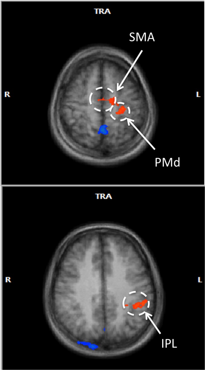
Motor imagery (MI) has shown effectiveness in enhancing motor performance. This may be due to the common neural mechanisms underlying MI and motor execution (ME). The main region of the ME network, the primary motor cortex (M1), has been consistently linked to motor performance. However, the activation of M1 during motor imagery is controversial, which may account for inconsistent rehabilitation therapy outcomes using MI. Here, we examined the relationship between contralateral M1 (cM1) activation during MI and changes in sensorimotor performance. To aid cM1 activity modulation during MI, we used real-time fMRI neurofeedback-guided MI based on cM1 hand area blood oxygen level dependent (BOLD) signal in healthy subjects, performing kinesthetic MI of pinching. We used multiple regression analysis to examine the correlation between cM1 BOLD signal and changes in motor performance during an isometric pinching task of those subjects who were able to activate cM1 during motor imagery. Activities in premotor and parietal regions were used as covariates. We found that cM1 activity was positively correlated to improvements in accuracy as well as overall performance improvements, whereas other regions in the sensorimotor network were not. The association between cM1 activation during MI with performance changes indicates that subjects with stronger cM1 activation during MI may benefit more from MI training, with implications toward targeted neurotherapy.

Keywords: motor imagery; motor skill; neurofeedback; real-time fMRI.


Figures

Figure 1
Figure 1
Experimental protocol and setup. (A) Structure of the rtfMRI session (see Methods). (B) Custom-built MR-compatible precision grip sensor used to perform precision grip both in the localizer and the isometric force target matching task, (C) Isometric force matching task in which the applied force (bar) has to match a horizontal line representing the target force (10 or 20% of MVF), (D) cM1 knob region (red) activated during the functional localizer, (E) Visual feedback displaying task instructions and a ball moving vertically during MI, proportional to the cM1 BOLD signal.
Figure 2
Figure 2
Inclusion criteria for successful trials during the force matching task. The two inclusion criteria for successful trials were target error (IE) being within 15% of target force (gray shaded area) and the first derivative of force () reaching 10% of maximum speed between 0.15 and 0.5 s after the visual cue (gray arrows). This figure shows two successive trials, the first trial (t = 0 s) fits both criteria, while the second one (t = 3.9 s) does not satisfy either criterion.
Figure 3
Figure 3
cM1 beta values in all eleven Participants (P1… P11). Individual cM1 beta values during baseline imagery, the three neurofeedback runs (NF run1, run2, run3) and the average across runs (Avg NF runs).
Figure 4
Figure 4
Correlations of cM1 up-regulation with behavioral outcome measures. Correlation of normalized cM1 beta values during neurofeedback-guided motor imagery with an overall improvement in motor performance (ΔMP, top), with a decrease in initial error (ΔIE, inverse of accuracy, middle) and a decreasing trend in maximum first force derivative (Δmax, speed of moving bar, bottom).
Figure 5
Figure 5
Voxel-wise RFX analysis of neurofeedback-guided motor imagery. Above, frontal lobe (z = 55 mm), below, inferior parietal lobule (z = 34 mm). Radiological convention (contralateral/left is on right). Cluster level corrected, p < 0.05. Orange: BOLD signal increase; blue: BOLD signal decrease.

Similar articles

Cited by

References

    1. Alkadhi H., Brugger P., Boendermaker S. H., Crelier G., Curt A., Hepp-Reymond M.-C., et al. . (2005). What disconnection tells about motor imagery: evidence from paraplegic patients. Cereb. Cortex 15, 131–140. 10.1093/cercor/bhh116 - DOI - PubMed
    1. Berman B. D., Horovitz S. G., Venkataraman G., Hallett M. (2012). Self-modulation of primary motor cortex activity with motor and motor imagery tasks using real-time fMRI-based neurofeedback. Neuroimage 59, 917–925. 10.1016/j.neuroimage.2011.07.035 - DOI - PMC - PubMed
    1. Binkofski F., Amunts K., Stephan K. M., Posse S., Schormann T., Freund H., et al. . (2000). Broca's region subserves imagery of motion: a combined cytoarchitectonic and fMRI study. Hum. Brain Mapp. 11, 273–285. 10.1002/1097-0193(200012)11:4%3C273::AID-HBM40%3E3.0.CO;2-0 - DOI - PMC - PubMed
    1. Bode S., Koeneke S., Jäncke L. (2007). Different strategies do not moderate primary motor cortex involvement in mental rotation: a TMS study. Behav. Brain Funct. 3:38. 10.1186/1744-9081-3-38 - DOI - PMC - PubMed
    1. Boecker H., Ceballos-Baumann A. O., Bartenstein P., Dagher A., Forster K., Haslinger B., et al. (2002). A H215O positron emission tomography study on mental imagery of movement sequences–the effect of modulating sequence length and direction. Neuroimage 17, 999–1009 10.1006/nimg.2002.1139 - DOI - PubMed

LinkOut - more resources