. 2015 Mar 12;11(3):e1004726.
doi: 10.1371/journal.ppat.1004726. eCollection 2015 Mar.

TRIM26 negatively regulates interferon-β production and antiviral response through polyubiquitination and degradation of nuclear IRF3

Affiliations

TRIM26 negatively regulates interferon-β production and antiviral response through polyubiquitination and degradation of nuclear IRF3

Peng Wang et al. PLoS Pathog. .

Abstract

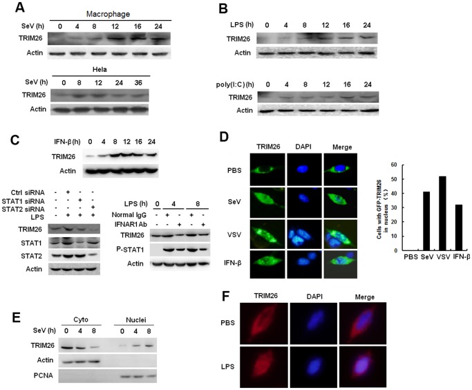
Virus infection leads to the activation of transcription factor IRF3 and subsequent production of type I inteferons, which induce the transcription of various antiviral genes called interferon stimulated genes (ISGs) to eliminate viral infection. IRF3 activation requires phosphorylation, dimerization and nuclear translocation. However, the mechanisms for the termination of IRF3 activation in nucleus are elusive. Here we report the identification of TRIM26 to negatively regulate IFN-β production and antiviral response by targeting nuclear IRF3. TRIM26 bound to IRF3 and promoted its K48-linked polyubiquitination and degradation in nucleus. TRIM26 degraded WT IRF3 and the constitutive active mutant IRF3 5D, but not the phosphorylation deficient mutant IRF3 5A. Furthermore, IRF3 mutant in the Nuclear Localization Signal (NLS), which could not move into nucleus, was not degraded by TRIM26. Importantly, virus infection promoted TRIM26 nuclear translocation, which was required for IRF3 degradation. As a consequence, TRIM26 attenuated IFN-β promoter activation and IFN-β production downstream of TLR3/4, RLR and DNA sensing pathways. TRIM26 transgenic mice showed much less IRF3 activation and IFN-β production, while increased virus replication. Our findings delineate a novel mechanism for the termination of IRF3 activation in nucleus through TRIM26-mediated IRF3 ubiquitination and degradation.


Conflict of interest statement

The authors have declared that no competing interests exist.

Figures

Fig 1
Fig 1. TRIM26 negatively regulates IFN-β production and antiviral response.
(A) RAW264.7, HEK293/TLR4 and HEK293/TLR3 cells were transfected with IFN-β reporter plasmid together with TRIM26 expression plasmid or control plasmid, analyzed luciferase activity after treatment with LPS and poly(I:C), respectively. (B) HEK293 cells were transfected with IFN-β reporter plasmid together with TRIM26 expression plasmid or control plasmid, analyzed luciferase activity after infection with SeV or transfection with poly(I:C). (C) Hela cells were transfected with IFN-β reporter plasmid together with TRIM26 expression plasmid or control plasmid, analyzed luciferase activity after transfection with ISD, poly(dA:dT) and cGAS expression plasmid. (D) Western blot analysis of TRIM26 expression in mouse peritoneal macrophages transfected with control siRNA, TRIM26 siRNA 1 or siRNA 2 for 36 h. (E–F) ELISA analysis of IFN-β production in mice peritoneal macrophages transfected with TRIM26 siRNA as in (D) followed stimulation with LPS, poly(I:C), SeV or ISD. (G) Hela cells (2×10⁵) were transfected with the TRIM26 expression plasmid or control plasmid and then infected with VSV (MOI, 0.1). Supernatants were analyzed for VSV titers with standard plaque assays. Intracellular VSV RNA replicates and IFN-β expression were measured by QRT-PCR. (H) Mouse primary peritoneal macrophages were transfected with control siRNA (Ctrl) or TRIM26 siRNA (siRNA) and then infected with VSV (MOI, 0.1). VSV titers, intracellular VSV RNA replicates and IFN-β expression were measured as in (G). Data are representative of three independent experiments (mean ± S.D. of quadruplicates in A-C and triplicates in E–H).
Fig 2
Fig 2. TRIM26 targets IRF3.
(A) HEK293/TLR3 and HEK293/TLR4 or Hela cells were transfected with ISRE reporter plasmid together with TRIM26 expression plasmid or control plasmid, analyzed luciferase activity after treatment with indicated agonists. (B) Western blot analysis of phosphorylated-IRF3 and total IRF3 in HEK293 cells transfected with TRIM26 expression plasmid or control plasmid, along with indicated adaptors. (C-D) Western blot analysis of phosphorylated-IRF3, and total IRF3 in mouse peritoneal macrophages transfected with control siRNA (Ctrl) or TRIM26 siRNA 1 (siRNA) and stimulated with LPS (C) or infected with SeV (D) for indicated times. (E) HEK293 cells were transfected with expression plasmids for TRIF, RIG-I, MAVS, TBK1 or IRF3 5D, along with IFN-β reporter plasmid and increasing amount of TRIM26 plasmid, and analyzed luciferase activity. (F) HEK293 cells were transfected with expression plasmids for RIG-I, MAVS, TRIF, TBK1 or IRF3 5D, along with ISRE reporter plasmid and TRIM26 plasmid, and analyzed luciferase activity. Data are representative of at least three independent experiments (mean ± S.D. of quadruplicates in A, E, F).
Fig 3
Fig 3. TRIM26 promotes IRF3 ubiquitination and proteasomal degradation.
(A) Lysates from mice peritoneal macrophages stimulated with LPS or infected with SeV for indicated time periods were subjected to immunoprecipitation with anti-IRF3 antibody followed by western blot analysis with anti-TRIM26 antibody. (B) Lysates from HEK293 cells transfected with Flag-TRIM26 and IRF3 WT, 5D, 5A or control vector and treated with MG-132 were subjected to immunoprecipitation with anti-Myc antibody followed by western blot analysis with anti-Flag antibody. (C) Lysates from HEK293 cells transfected with expression plasmids for Myc-IRF3, Flag-TRIM26 WT, C16A, C16/36A and HA-Ub plasmids were subjected to immunoprecipitation with anti-Myc antibody followed by western blot analysis with anti-Ub antibody. (D) TRIM26 WT, C16/36A and WT IRF3 were obtained by in vitro transcription and translation. Interaction between TRIM26 and IRF3 was assayed by mixing WT TRIM26 and WT IRF3 together followed by IP with TRIM26 antibody and WB with IRF3 antibody. In vitro ubiquitination assay was performed in the presence of Ub, E1, UbcH5c, TRIM26 and IRF3. The ubiquitination of IRF3 was examined by WB with IRF3 antibody. (E) Lysates from mouse peritoneal macrophages transfected with control siRNA (Ctrl) or TRIM26 siRNA (siRNA) pretreated with MG-132 for 2 h followed with stimulation with LPS or infection with SeV for indicated time periods were subjected to immunoprecipitation with anti-IRF3 antibody followed by western blot analysis with anti-Ub antibody. (F) Lysates from HEK293 cells transiently transfected with expression plasmids for Myc-IRF3, Flag-TRIM26 and HA-Ub (WT), HA-Ub (K48) or HA-Ub (K63) were subjected to immunoprecipitation with anti-Myc antibody followed by western blot analysis with anti-Ub or anti-HA antibody. (G) Western blot analysis of Myc-IRF3 expression in HEK293 cells transfected with expression plasmids for Myc-IRF3, Flag-TRIM26 WT or TRIM26 C16A and then treated with MG132 or Chloroquine for 4 h. (H) Lysates from HEK293 cells transiently cotransfected with expression plasmids for Myc-IRF3 WT or K70/87R, along with Flag-TRIM26 and HA-Ub plasmids were subjected to immunoprecipitation with anti-Myc antibody followed by western blot analysis with anti-Ub or anti-Ub(K48) antibody. Similar results were obtained from three independent experiments.
Fig 4
Fig 4. Virus infection induces TRIM26 expression and nuclear translocation.
(A) Western blot analysis of TRIM26 protein expression in mouse primary peritoneal macrophages and Hela cells infected with SeV for indicated times. (B) Western blot analysis of TRIM26 protein expression in mouse primary peritoneal macrophages stimulated with LPS or poly(I:C) for indicated times. (C) Western blot analysis of TRIM26 protein expression in mouse peritoneal macrophages treated with mIFN-β for indicated times (left). Western blot analysis of TRIM26 protein expression in mouse peritoneal macrophages transfected with STAT1 siRNA, STAT2 siRNA and control siRNA (right, upper panel), or pretreated with IFNR1 antibody and control antibody for 2 h (right, lower panel), then simulated with LPS for indicated times. (D) Fluorescent images of HEK293 cells transfected with GFP-TRIM26 and then stimulated with SeV (MOI 1), VSV (MOI 0.1) or IFN-β for 8 h. Nuclei were detected with DAPI (blue). (E) Western blot analysis of TRIM26 protein expression in nuclear and cytoplasmic fractions from Hela cells infected with SeV for 4 and 8 h. (F) Immunofluorescent images of mouse peritoneal macrophages stimulated with 100 ng/ml LPS for 10 h. Endogenous TRIM26 was analyzed by immunostaining with anti-TRIM26 antibody (red). Similar results were obtained from three independent experiments.
Fig 5
Fig 5. TRIM26 promotes IRF3 degradation in nucleus.
(A) Nuclear and cytoplasmic fractions from RAW264.7 macrophages infected with SeV for 8 h or left uninfected were subjected to immunoprecipitation with anti-IRF3 antibody followed by western blot analysis with anti-TRIM26 antibody. (B) Nuclear and cytoplasmic fractions from RAW264.7 macrophages transfected with control siRNA (Ctrl) or TRIM26 siRNA (siRNA) and infected with SeV were subjected to immunoprecipitation with anti-IRF3 antibody followed by western blot analysis with anti-Ub antibody. (C) Western blot analysis of IRF3 expression in HEK293 cells transfected with expression plasmids for Myc-IRF3 WT, 5A or 5D and increased amount of Flag-TRIM26. (D) Western blot analysis of IRF3 expression in the cytoplasmic and nuclear fractions prepared from HEK293 cells, which were transfected with Myc-IRF3 WT and Flag-TRIM26 or control vector followed with SeV infection. Similar results were obtained from three independent experiments.
Fig 6
Fig 6. Nuclear translocation promotes TRIM26-mediated IRF3 degradation.
(A) Western blot analysis of IRF3 expression in HEK293 cells transfected with expression plasmids for Myc-IRF3 WT, KR77/78NG, 5D or 5D KR77/78NG and Flag-TRIM26. (B) Western blot analysis of IRF3 IL139/140MM expression in the cytoplasmic and nuclear fractions prepared from HEK293 cells, which were transfected with Myc-IRF3 IL139/140MM and Flag-TRIM26 or control vector followed with SeV infection. (C) Fluorescent images of HEK293 cells transfected with GFP-TRIM26 or GFP-TRIM26 NLS plasmid and infected with SeV and VSV. Nuclei were detected with DAPI (blue). (D) Western blot analysis of IRF3 expression in HEK293 cells transfected with expression plasmids for Myc-IRF3 WT, 5A or 5D and GFP-TRIM26 or GFP-TRIM26 NLS. (E) Western blot analysis of IRF3 expression in HEK293 cells transfected with Myc-IRF3 and Flag-TRIM26 pretreated with 20μM Ivermectin for 2 h. Similar results were obtained from three independent experiments.
Fig 7
Fig 7. Impaired IFN-β signaling and antiviral responses in TRIM26 transgenic mice.
(A–B) Expression of IFN-β mRNA and secretion of IFN-β protein measured by QRT-PCR and ELISA, respectively, in peritoneal macrophages from TRIM26-Tg and WT mice stimulated with LPS and poly(I:C) or infected with SeV and VSV for indicated times. (C) Peritoneal macrophages from TRIM26-Tg and WT mice were infected with VSV (MOI, 0.1) for 12 h. Supernatants were analyzed for VSV titers with standard plaque assays. (D) ELISA analyses of IFN-β production in sera, lung or liver from TRIM26-Tg and WT mice intravenously infected with VSV for 72 h. (n = 4 mice per group). (E) TRIM26-Tg and WT mice were infected with VSV as in D. VSV titers in lung and liver were determined by standard plaque assays (right). Expression of VSV G protein was analyzed by western blot with VSV-G antibody (left). (F) Hematoxylin and eosin staining of lung tissue sections from TRIM26-Tg and WT mice infected with VSV as in e. (G) Survival of TRIM26-Tg and WT mice (n = 10) infected with VSV (4×10⁷ pfu/mouse). Data are representative of three independent experiments (mean ± S.D. of triplicates in A-D).

References

    1. Janeway CA Jr, Medzhitov R (2002) Innate immune recognition. Annu Rev Immunol 20: 197–216. - PubMed
    1. Takeuchi O, Akira S (2010) Pattern recognition receptors and inflammation. Cell 140: 805–820. 10.1016/j.cell.2010.01.022 - DOI - PubMed
    1. Yoneyama M, Kikuchi M, Natsukawa T, Shinobu N, Imaizumi T, et al. (2004) The RNA helicase RIG-I has an essential function in double-stranded RNA-induced innate antiviral responses. Nat Immunol 5: 730–737. - PubMed
    1. Kato H, Takeuchi O, Sato S, Yoneyama M, Yamamoto M, et al. (2006) Differential roles of MDA5 and RIG-I helicases in the recognition of RNA viruses. Nature 441: 101–105. - PubMed
    1. Sun L, Wu J, Du F, Chen X, Chen ZJ (2013) Cyclic GMP-AMP synthase is a cytosolic DNA sensor that activates the type I interferon pathway. Science 339: 786–791. 10.1126/science.1232458 - DOI - PMC - PubMed

Publication types