. 2015 Mar 10;16(3):5434-51.
doi: 10.3390/ijms16035434.

SOCS3 silencing attenuates eosinophil functions in asthma patients

Affiliations

SOCS3 silencing attenuates eosinophil functions in asthma patients

Ma Paz Zafra et al. Int J Mol Sci. .

Abstract

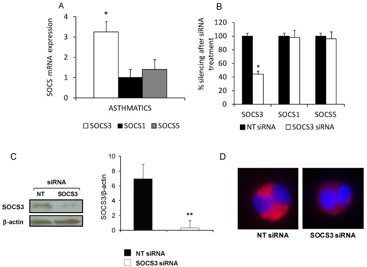
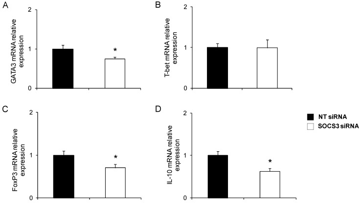
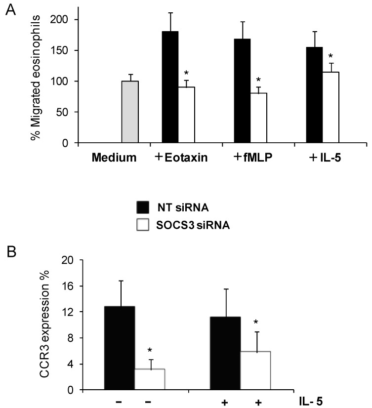
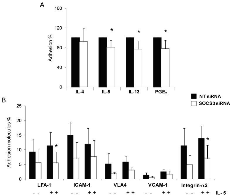
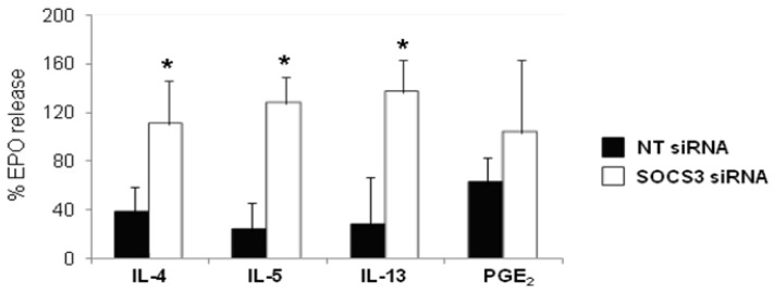
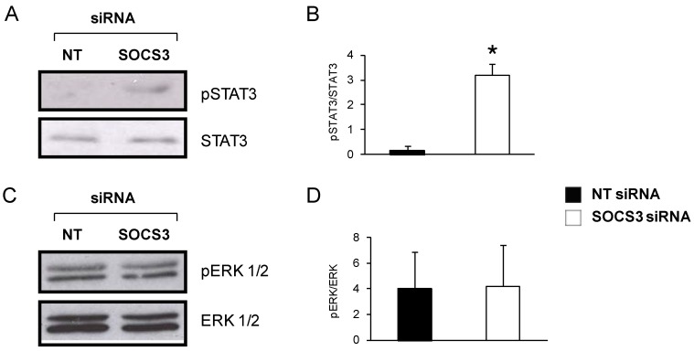
Eosinophils are one of the key inflammatory cells in asthma. Eosinophils can exert a wide variety of actions through expression and secretion of multiple molecules. Previously, we have demonstrated that eosinophils purified from peripheral blood from asthma patients express high levels of suppressor of cytokine signaling 3 (SOCS3). In this article, SOCS3 gene silencing in eosinophils from asthmatics has been carried out to achieve a better understanding of the suppressor function in eosinophils. SOCS3 siRNA treatment drastically reduced SOCS3 expression in eosinophils, leading to an inhibition of the regulatory transcription factors GATA-3 and FoxP3, also interleukin (IL)-10; in turn, an increased STAT3 phosphorilation was observed. Moreover, SOCS3 abrogation in eosinophils produced impaired migration, adhesion and degranulation. Therefore, SOCS3 might be regarded as an important regulator implicated in eosinophil mobilization from the bone marrow to the lungs during the asthmatic process.


Figures

Figure 1
Figure 1
siRNA technology reduces suppressor of cytokine signaling 3 (SOCS3) expression in eosinophils from asthmatic patients. (A) SOCS3, SOCS1 and SOCS5 mRNA relative levels were measured by quantitative PCR in eosinophils purified from asthmatic patients (n = 36); (B) After SOCS3 siRNA incubation, quantitative-PCR results showed significant down-regulation of SOCS3 mRNA levels and no alteration of SOCS5 or SOCS1, as well as decreased protein expression level of SOCS3 after SOCS3 siRNA treatment was detected by Western blot analysis and further quantified by densitometry (C) or immnunofluoresce (D) as is depicted on a representative picture of an eosinophil incubated with non-target (NT)siRNA (left panel) or SOCS3 siRNA (right panel), after staining with anti-SOCS3 antibody labeled with Alexa 647 fluorochrome. The results were expressed as mean ± SD, (n = 6–8). * Denotes statistical significance (p < 0.05); ** denotes obvious statistical significance (p < 0.01).
Figure 2
Figure 2
Phenotype evaluation in SOCS3-deficient eosinophils purified from asthma patients. GATA3 (A); T-bet (B); FoxP3 (C); and IL-10 (D) mRNA relative levels were determined by qPCR in blood eosinophils purified from five asthma patients treated with SOCS3 siRNA or NT siRNA. * p < 0.05 between groups.
Figure 3
Figure 3
Reduced eosinophil migration due to SOCS3 down-regulation by gene silencing. Cell migration was detected by a transwell assay; percentages of eosinophils migrated towards IL-5, eotaxin and fMLP (A) were measured by flow cytometry after SOCS3 silencing; and (B) CCR3 surface expression on eosinophils was achieved by the measure of FITC positive cells by flow cytometry. The results were expressed as mean ± SD, (n = 6–8). * Denotes statistical significance (p < 0.05) between groups.
Figure 4
Figure 4
Impaired adhesion capacity in blood eosinophils from asthmatic patients after SOCS3 gene silencing. (A) NT and SOCS3 siRNA treated eosinophils were incubated with IL-4, IL-5, IL-13, and PGE2 for 1 or 2 h and then allowed to adhere to fibronectin-coated wells for 30 min. Results are expressed as mean ± SD of adhered cell percentages estimated by residual eosinophils peroxidase (EPO) measurement; and (B) LFA-1, ICAM-1, VLA4, VCAM-1, and Integrin-α2 surface expression on blood eosinophils from seven asthmatic patients incubated with SOCS3 siRNA or NT siRNA were determined by flow cytometry. * p < 0.05 between groups.
Figure 5
Figure 5
Augmented eosinophil degranulation in asthmatic blood eosinophils treated with SOCS3 siRNA. Purified blood eosinophils from eight asthmatic patients incubated with SOCS3 siRNA (white bars, mean ± SD) or NT siRNA (black bars, mean ± SD) for 48 h, were pretreated with IL-4, IL-5, IL-13 and PGE2 for 1 or 2 h and them stimulated with C5a (300 nM) for 30 min at 37 °C. The release of EPO activity into supernatants was determined by photometry. Data is expressed as a percentage of the maximal control response (300 nM). * p < 0.05 between groups.
Figure 6
Figure 6
Determination of pSTAT3 and pERK 1/2 protein levels in eosinophils from asthmatic patients. (A) Representative pSTAT3 and STAT3 Western blots of blood eosinophils from an asthmatic patient treated with SOCS3 siRNA or NT siRNA for 48 h; (B) Densitometric quantification expressed as mean ± SD, n = 3. * p < 0.05 between groups; (C) Eosinophil pERK 1/2 and ERK Western blot after treatment with SOCS3 siRNA or NT siRNA for 48 h; and (D) pERK 1/2 and ERK bands were quantified by densitometry. SOCS3 siRNA (white bars, mean ± SD) and NT siRNA (black bars, mean ± SD), n = 3.

References

    1. Woodruff P.G., Modrek B., Choy D.F., Jia G., Abbas A.R., Ellwanger A., Koth L.L., Arron J.R., Fahy J.V. T-helper type 2-driven inflammation defines major subphenotypes of asthma. Am. J. Respir. Crit. Care Med. 2009;180:388–395. doi: 10.1164/rccm.200903-0392OC. - DOI - PMC - PubMed
    1. Rothenberg M.E., Hogan S.P. The eosinophil. Annu. Rev. Immunol. 2006;24:147–174. doi: 10.1146/annurev.immunol.24.021605.090720. - DOI - PubMed
    1. Rosenberg H.F., Phipps S., Foster P.S. Eosinophil trafficking in allergy and asthma. J. Allergy Clin. Immunol. 2007;119:1303–1310. doi: 10.1016/j.jaci.2007.03.048. - DOI - PubMed
    1. Akuthota P., Wang H.B., Spencer L.A., Weller P.F. Immunoregulatory roles of eosinophils: A new look at a familiar cell. Clin. Exp. Allergy. 2008;38:1254–1263. doi: 10.1111/j.1365-2222.2008.03037.x. - DOI - PMC - PubMed
    1. Woerly G., Roger N., Loiseau S., Capron M. Expression of Th1 and Th2 immunoregulatory cytokines by human eosinophils. Int. Arch. Allergy Immunol. 1999;118:95–97. doi: 10.1159/000024038. - DOI - PubMed

Publication types

MeSH terms

Substances