Review
. 2014 Oct-Dec;29(4):241-53.

Magnetoencephalography in the study of brain dynamics

Review

Magnetoencephalography in the study of brain dynamics

Vittorio Pizzella et al. Funct Neurol. 2014 Oct-Dec.

Abstract

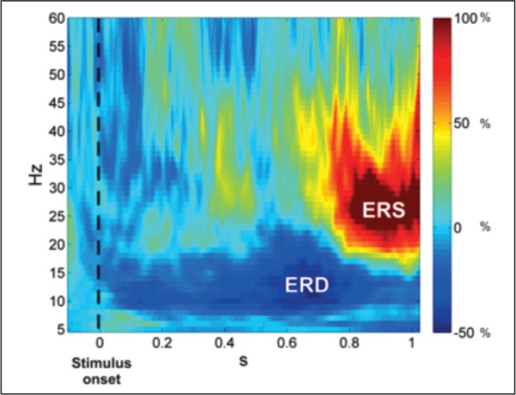
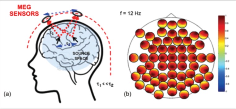
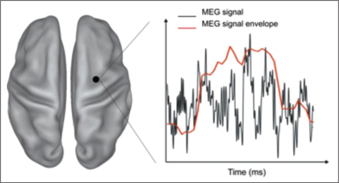
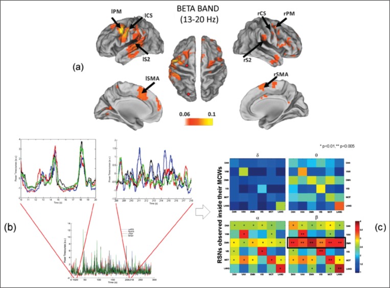
To progress toward understanding of the mechanisms underlying the functional organization of the human brain, either a bottom-up or a top-down approach may be adopted. The former starts from the study of the detailed functioning of a small number of neuronal assemblies, while the latter tries to decode brain functioning by considering the brain as a whole. This review discusses the top-down approach and the use of magnetoencephalography (MEG) to describe global brain properties. The main idea behind this approach is that the concurrence of several areas is required for the brain to instantiate a specific behavior/functioning. A central issue is therefore the study of brain functional connectivity and the concept of brain networks as ensembles of distant brain areas that preferentially exchange information. Importantly, the human brain is a dynamic device, and MEG is ideally suited to investigate phenomena on behaviorally relevant timescales, also offering the possibility of capturing behaviorally-related brain connectivity dynamics.


Figures

None
(a) Post-synaptic activity of a single neuron can be effectively modeled by a single current dipole (orange arrow); (b) when many pyramidal neurons are synchronously active and spatially well aligned, an equivalent current dipole may be used to model cortical activity (red arrow); (c) if a spherical model for the head is used, only dipoles located in the fissures generate a magnetic field outside the head.
None
The 153-channel MEG system installed at ITAB -University of Chieti. It consists of 153 channels arranged on a helmet surface, so as to simultaneously record the brain magnetic field at multiple sampling points. Each channel is based on superconducting devices acting as magnetic field sensors. The sensor array is contained in a non-magnetic cryostat. The whole system is installed in a high quality magnetically shielded room.
None
(a) Individual MRI data and superimposed results of tissue boundary segmentation (yellow contours). (b) Realistic volume conductor head model reconstructed from the geometric tessellation of the head tissue. (c) Source and volume conductor model are used to identify brain sources from MEG data. Images derived from the Curry 6.0 (Neuroscan) analysis software.
None
Neuronal oscillations can be observed at different spatial scales: from single units, to local field potential, intracranial EEG, and MEG (modified from Varela et al., 2001). Oscillations generated by a large amount of cells, like those recorded by scalp EEG or MEG, have a spectral content that can be divided into different bands. Modified from Varela et al. (2001).
Figure 1
Figure 1
Event-related synchronization and desynchronization in the right secondary somatosensory area after galvanic stimulation. Power changes in the right secondary somatosensory area as a function of time (seconds) and frequency (Hz) relative to the baseline period (−0.1s–0s), zero being the onset of a galvanic stimulation delivered to the index of the left hand in a single right-handed subject). Blue indicates a power decrease, i.e. ERD, and red indicates a power increase (ERS). ERD/ERS phenomena show different latencies and spectral contents, with ERD in the alpha-frequency range (8–12 Hz) and ERS in the beta range (15–25 Hz).
Figure 2
Figure 2
Schematic representation of signal propagation from brain sources to MEG sensors (a) and of channel-level interactions for a subset of MEG channels (b). a) signal propagation to the sensors is assumed to be instantaneous in comparison to the timescales of signal propagation within the brain (τ1<<τ2); at a given time instant, the different MEG sensors capture a weighted sum of the activities of all brain sources. b) Schematic representation of channel-level interactions for a subset of MEG channels. The larger black circle indicates the system layout and each smaller circle indicates the coupling of one sensor (black dot) with all the others. The spread of the source activity to the sensors artificially enhances the degree of coupling between channels independently of the actual brain source interaction. Indeed, in this toy example all channels appear to be highly coupled with all the others although only two interacting sources were simulated (a).
Figure 3
Figure 3
Dynamic information of the MEG signal. MEG signal estimated at one brain location (black dot) shows a rich temporal structure at the millisecond timescale (black curve). Fluctuations of signal envelope capture slowly varying MEG power dynamics (red curve) with a timescale similar to fMRI signal fluctuations.
Figure 4
Figure 4
Brain networks as observed by magnetoencephalography. (a) Phase coupling of band-limited oscillatory signals as identified by multivariate interaction measure between the dorsal attention network (DAN) and the motor network in the beta band. (b) Correlated fluctuations of band power envelopes of resting-state MEG show time-variant profiles, e.g., when the within-DAN connectivity is computed in two different temporal epochs. Time windows of stronger connectivity alternate with periods of lower connectivity. (c) Correlated fluctuations of band power envelope for various RSNs show different behaviors for different frequencies. Notably, the default mode network plays a central role in brain functional connectivity in the beta band. Modified from de Pasquale et al. (2012) and Marzetti et al. (2013).

References

    1. Ahlfors SP, Simpson GV, Dale AM, et al. Spatiotemporal activity of a cortical network for processing visual motion revealed by MEG and fMRI. J Neurophysiol. 1999;82:2545–2555. - PubMed
    1. Baillet S, Mosher JC, Leahy RM. Electromagnetic brain mapping. IEEE Signal Processing Magazine. 2001;18:14–30.
    1. Beggs JM, Plenz D. Neuronal avalanches in neocortical circuits. J Neurosci. 2003;23:11167–11177. - PMC - PubMed
    1. Betti V, Della Penna S, de Pasquale F, et al. Natural scenes viewing alters the dynamics of functional connectivity in the human brain. Neuron. 2013;79:782–797. - PMC - PubMed
    1. Biswal B, Yetkin FZ, Haughton VM, et al. Functional connectivity in the motor cortex of resting human brain using echo-planar MRI. Magn Reson Med. 1995;34:537–541. - PubMed

Publication types

LinkOut - more resources