Review
. 2014;5(3):e28674.
doi: 10.4161/trns.28674.

The Old and New Testaments of gene regulation. Evolution of multi-subunit RNA polymerases and co-evolution of eukaryote complexity with the RNAP II CTD

Affiliations
Review

The Old and New Testaments of gene regulation. Evolution of multi-subunit RNA polymerases and co-evolution of eukaryote complexity with the RNAP II CTD

Zachary F Burton. Transcription. 2014.

Abstract

I relate a story of genesis told from the point of view of multi-subunit RNA polymerases (RNAPs) including an Old Testament (core RNAP motifs in all cellular life) and a New Testament (the RNAP II heptad repeat carboxy terminal domain (CTD) and CTD interactome in eukarya). The Old Testament: at their active site, one class of eukaryotic interfering RNAP and ubiquitous multi-subunit RNAPs each have two-double psi β barrel (DPBB) motifs (a distinct pattern for compact 6-β sheet barrels). Between β sheets 2 and 3 of the β subunit type DPBB of all multi-subunit RNAPs is a sandwich barrel hybrid motif (SBHM) that interacts with conserved initiation and elongation factors required to utilize a DNA template. Analysis of RNAP core protein motifs, therefore, indicates that RNAP evolution can be traced from the RNA-protein world to LUCA (the last universal common ancestor) branching to LECA (the last eukaryotic common ancestor) and to the present day, spanning about 4 billion years. The New Testament: in the eukaryotic lineage, I posit that splitting RNAP functions into RNAPs I, II and III and innovations developed around the CTD heptad repeat of RNAP II and the extensive CTD interactome helps to describe how greater structural, cell cycle, epigenetic and signaling complexity co-evolved in eukaryotes relative to eubacteria and archaea.

Keywords: Chromatin and transcription; Eukaryotic transcription; Prokaryotic transcription; RNA polymerases; transcriptional elongation; transcriptional initiation; transcriptional termination.


Figures
None
Figure 1. The active site of eubacterial Tt RNAP. The catalytic center is occupied by ATP-Mg (space-filling representation). β sheets are yellow. RNA is silver. DNA is gold (template strand) and green (non-template strand). The active site is wedged between the closed trigger loop (TL; silver), bridge helix (BH; orange) and two DPBBs. This image was drawn using PDB 205J with the program Visual Molecular Dynamics (used for all molecular graphics).
None
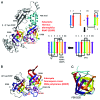
Figure 2. Two-DPBB type DDRPs and RDRPs. (A) Tt RNAP DPBBs. (B) Neurospora crassa interfering RNA polymerase (RDRP) DPBBs. (C) A simpler 6-β sheet barrel from translation elongation factor EF-G from Staphylococcus aureus from which a DPBB can be derived via β sheet exchange (β2 for β5). The schematic indicates a potential two step evolution of DPBBs by duplication of a simple 3-sheet motif to form a simple 6-sheet barrel (as in EF-G) followed by switching the order of β2 and β5. The color coding for the β sheets is indicated, and sheets are numbered β1→β6 according to their order in the peptide chain. The Greek letter psi (ψ) indicates the two psi pattern in the DPBB. Connections in the β type DPBB are silver; connections in the β’ type DPBB are cyan; Mg is magenta. Active site aspartates are shown. Small silver arrows emphasize a conserved loop (Mg binding) and motif between β5 and β6.
None
Figure 3. A SBHM necessary to utilize a DNA template. The SBHM is inserted between β2 and β3 of the β subunit type DPBB and is a landing pad for conserved initiation and elongation factors in the three domains of cellular life: eubacteria (red), archaea (cyan), eukarya (purple). Coloring of the DPBBs is as in Figure 2.
None
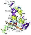
Figure 4. Multi-subunit RNAPs from eukarya and eubacteria are closely related. (A) Sc RNAP II (eukaryotic). (B) Tt RNAP (eubacterial). DPBBs are colored as in Figure 2 (see schematic). Only a subset of core motifs is shown. RNA is red; DNA is blue (template strand) or green (non-template strand). The bridge helix (BH) is magenta; the trigger loop (TL) is ice blue.
None
Figure 5. A rough timeline and description of the evolution of two DPBB type DDRPs and related RDRPs and proposed mechanisms for innovation in evolution.
None
Figure 6. The Old and New Testaments of molecular biology describing evolution of multi-subunit RNAPs from the RNA-protein world to LUCA and branching to LECA. The red arrows indicate that LUCA may have eaten the RNA-protein world and that more modern organisms may have devoured and/or out competed LUCA. According to this description, eubacteria and archaea maintain similar features to LUCA. Higher order complexity in eukaryotic gene regulation developed around the CTD of RNAP II and chromatin, and these processes are posited to be strongly co-evolved.
None
Figure 7. The CTD of RNAP II is partly analogous to a GPS for the transcription cycle. The RNAP II transcription cycle, which is centered on the CTD and its interactome, appears co-evolved with the eukaryotic cell cycle. The sequence of the 52 heptad repeat human CTD is shown. Non-consensus heptads are indicated by red letters.
None
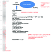
Figure 8. Gene regulatory network (GRN) structures (blue arrows) and co-evolved processes (green arrows) for (A) eukaryotes and (B) eubacteria.

References

    1. Wang B, Feig M, Cukier RI, Burton ZF. Computational simulation strategies for analysis of multisubunit RNA polymerases. Chem Rev. 2013;113:8546–66. doi: 10.1021/cr400046x. - DOI - PMC - PubMed
    1. Werner F. Molecular mechanisms of transcription elongation in archaea. Chem Rev. 2013;113:8331–49. doi: 10.1021/cr4002325. - DOI - PubMed
    1. Weinzierl RO. The RNA polymerase factory and archaeal transcription. Chem Rev. 2013;113:8350–76. doi: 10.1021/cr400148k. - DOI - PubMed
    1. Iyer LM, Koonin EV, Aravind L. Evolutionary connection between the catalytic subunits of DNA-dependent RNA polymerases and eukaryotic RNA-dependent RNA polymerases and the origin of RNA polymerases. BMC Struct Biol. 2003;3:1. doi: 10.1186/1472-6807-3-1. - DOI - PMC - PubMed
    1. Lane WJ, Darst SA. Molecular evolution of multisubunit RNA polymerases: structural analysis. J Mol Biol. 2010;395:686–704. doi: 10.1016/j.jmb.2009.10.063. - DOI - PMC - PubMed

Publication types

LinkOut - more resources