. 2015 Mar 12;13(3):e1002089.
doi: 10.1371/journal.pbio.1002089. eCollection 2015 Mar.

ATPase-dependent control of the Mms21 SUMO ligase during DNA repair

Affiliations

ATPase-dependent control of the Mms21 SUMO ligase during DNA repair

Marcelino Bermúdez-López et al. PLoS Biol. .

Abstract

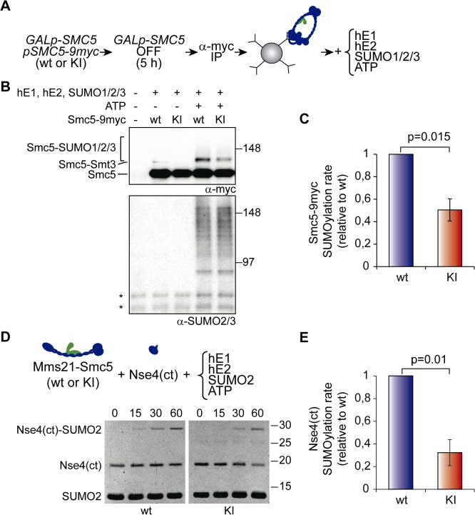
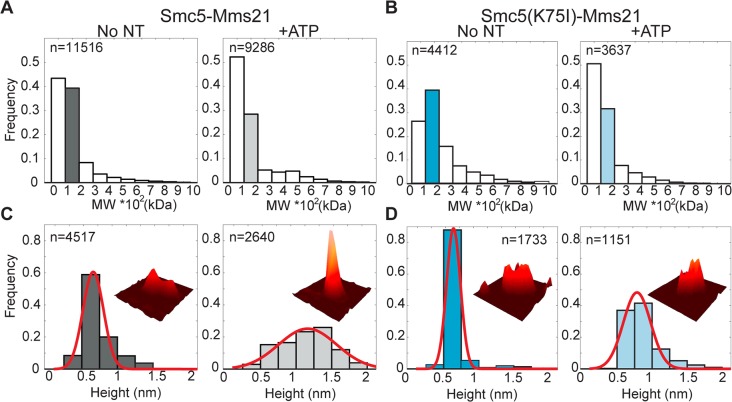
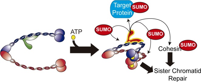
Modification of proteins by SUMO is essential for the maintenance of genome integrity. During DNA replication, the Mms21-branch of the SUMO pathway counteracts recombination intermediates at damaged replication forks, thus facilitating sister chromatid disjunction. The Mms21 SUMO ligase docks to the arm region of the Smc5 protein in the Smc5/6 complex; together, they cooperate during recombinational DNA repair. Yet how the activity of the SUMO ligase is controlled remains unknown. Here we show that the SUMO ligase and the chromosome disjunction functions of Mms21 depend on its docking to an intact and active Smc5/6 complex, indicating that the Smc5/6-Mms21 complex operates as a large SUMO ligase in vivo. In spite of the physical distance separating the E3 and the nucleotide-binding domains in Smc5/6, Mms21-dependent sumoylation requires binding of ATP to Smc5, a step that is part of the ligase mechanism that assists Ubc9 function. The communication is enabled by the presence of a conserved disruption in the coiled coil domain of Smc5, pointing to potential conformational changes for SUMO ligase activation. In accordance, scanning force microscopy of the Smc5-Mms21 heterodimer shows that the molecule is physically remodeled in an ATP-dependent manner. Our results demonstrate that the ATP-binding activity of the Smc5/6 complex is coordinated with its SUMO ligase, through the coiled coil domain of Smc5 and the physical remodeling of the molecule, to promote sumoylation and chromosome disjunction during DNA repair.


Conflict of interest statement

The authors have declared that no competing interests exist.

Figures

Fig 1
Fig 1. The Smc5-Mms21 interaction is required for sumoylation of Mms21 targets and chromosome segregation after DNA damage.
A. Models for Mms21-dependent sumoylation: Mms21 may target proteins (including cohesin subunits) from its location in the Smc5/6 complex (left), or independently from Smc5/6 (right). B. Scheme of Mms21-binding surface on the coiled coil 2 of the Smc5 protein and location of mutated sites. C. Co-immunoprecipitation analysis of the Smc5-Mms21 and Smc5-Nse4 interactions. MMS21-6HA and NSE4-6HA cells were transformed with centromeric plasmids expressing the indicated SMC5 alleles and subjected to anti-HA immunoprecipitation. D. Growth test analysis of GALp-SMC5 cells transformed with the indicated centromeric plasmids. E and F. GALp-SMC5 cells bearing the indicated vectors were shifted to glucose for 4 h to repress expression of the endogenous SMC5 gene, and then treated as depicted in the figure; samples were taken at the indicated times for 4’,6-diamidino-2-phenylindole (DAPI) staining and microscopic examination (E) or Fluorescence-Activated Cell Sorting (FACS) analysis (F). Rectangles in F mark cells with less than 1N DNA content. G. GALp-SMC5 cells ectopically expressing the indicated SMC5 alleles from a centromeric vector were shifted to glucose for 6 h; 6xHis-Flag (HF) tagged SUMO was pulled down (P.D.) under denaturing conditions from yeast protein extracts (Input) to purify sumoylated species. Input and P.D. samples were analyzed by western blot with the indicated antibodies. H. GALp-SMC5 SMC1-6HA cells expressing the indicated SMC5 alleles from a plasmid were shifted to glucose for 6 h. Protein extracts were processed for SUMO pull-down analysis as in G to analyze Smc1 sumoylation. In C, G, and H: wt = wild type, S1 = smc5-S1, S2 = smc5-S2, S3 = smc5-S3. In G and H, arrow points to unmodified form of the proteins, vertical bar to sumoylated forms; re-probing with anti-Flag is shown as a loading control for total SUMO in the purification.
Fig 2
Fig 2. Mms21-dependent sumoylation requires an intact Smc5/6 complex.
A. Composition of Smc5/6, depicting the different entities present in the complex. Nse subunits are labeled 1 to 6; Nse2 = Mms21. B. Sumoylation of Smc5 in smc6 mutant cells. Samples of wild type and GAL-3HA-SMC6 were collected from cells growing exponentially in galactose (GALp ON), or 12 h after shift to glucose to repress 3HA-SMC6 expression (GALp OFF). A GALp-3HA-SMC6 strain expressing the smc6-1 allele from a centromeric vector was also included in the analysis. Protein extracts were processed for HF-SUMO pull down as in Fig. 1G. C. Co-immunoprecipitation analysis of the Smc5-Mms21 interaction from wild type and smc6-1 protein extracts. Wild type and GALp-SMC6 smc6-1 cells expressing Smc5-9myc and Mms21-6HA were shifted to glucose for 12 h and processed for anti-HA immunoprecipitation. D. Chromatin fractionation assay from wild type and smc6-1 cells to analyze the amount of chromatin-bound Mms21-6HA. Controls for a chromatin-bound protein (histone H3) and cytoplasmic soluble (Hexokinase; Hxk) proteins are shown. E. Temperature and methyl methanesulfonate (MMS)-sensitivity of nse hypomorphic alleles. Growth test of wild type, nse3-2, and nse5-2 cells in YPD plates at 25°C (containing or not the indicated MMS concentration) or at 37°C. F. Analysis of the Smc5-Nse3 and Smc5-Nse5 interaction in nse hypomorphic alleles. Exponentially growing Smc5-6Flag cells, expressing 9myc-tagged versions of either the wild type or the indicated hypomorphic nse alleles, were shifted to 37°C for 2 h (37) or kept at 25°C (25) before Smc5-6Flag immunoprecipitation. G. Co-immunoprecipitation analysis of the Smc5-Mms21 interaction in nse3-2 and nse5-2 mutant cells. Smc5-6Flag was immunoprecipitated, as in F, from cells grown at the indicated temperatures. Co-immunoprecipitation of Mms21-6HA was analyzed by western blot. H. Chromatin fractionation assay from Mms21-6HA tagged wild type and nse5-2 cells, as in D. I. HF-SUMO pull down from wild type, nse3-2, or nse5-2 cells expressing Smc5-6HA, before and after a shift to 37°C. In B and G, arrow points to unmodified form of the proteins, vertical bar to sumoylated forms. In D and H, WCE: Whole Cell Extract; SN: Supernatant; Chr: Chromatin fraction.
Fig 3
Fig 3. ATPase-dependent activity of the Mms21 SUMO ligase.
A. Growth test of GALp-SMC5 cells expressing wild-type SMC5, smc5(K75I), or smc5(D1014A) from a centromeric vector in plates containing galactose (GALp ON) or glucose (GALp OFF). B. Mms21-3HA was immunoprecipitated from exponentially growing cells transformed with the indicated SMC5-expressing centromeric plasmids to test the Smc5-Mms21 interaction; wt = wild type; KI = smc5(K75I); DA = smc5(D1014A). C. Sumoylation analysis of ATPase-defective Smc5-9myc proteins. HF-SUMO pull-down analysis in wild-type cells transformed with plasmids expressing the indicated SMC5 alleles. D. Sumoylation analysis of Nse4-6HA in smc5 ATPase mutant cells. HF-SUMO pull-down analysis in GALp-SMC5 NSE4-6HA cells expressing the indicated SMC5 alleles. Cells were shifted to glucose 6 h before collection to repress the endogenous SMC5 gene. E. Sumoylation analysis of cohesin in smc5 ATPase mutant cells. HF-SUMO pull down from cells of the indicated genotype (wt, mms21ΔC and GALp-SMC5), carrying a C-terminal 6HA tag on SMC1, and expressing or not an ectopic copy of SMC5-9myc (WT) or smc5(K75I)-9myc (KI) allele; where indicated, cells were treated with MMS 0,02% for 1 h (MMS) before collection. F. Chromatin fractionation assay from GALp-SMC5 MMS21-6HA cells expressing an ectopic 9myc-tagged copy of the indicated SMC5 alleles, collected 6 h after shift to glucose to deplete the endogenous Smc5 protein. Controls for a chromatin-bound protein (histone H3), nuclear soluble (Rpd3) and cytoplasmic soluble (Hexokinase; Hxk) proteins are shown; WCE: Whole Cell Extract; SN: Supernatant; Chr: Chromatin fraction. In C–E, arrow points to unmodified Smc5, Nse4, or Smc1 proteins, and vertical bars to their sumoylated forms.
Fig 4
Fig 4. The ATPase in the Smc5/6 complex is part of the ligase mechanism that triggers sumoylation.
A. Outline of an Mms21-Ubc9 (E3-E2) fusion, using a 3xHA linker, to force constitutive Ubc9 recruitment in the vicinity of the SUMO ligase. B. Growth test analysis of fusions of the E2 to a full-length Mms21 or to an mms21Δc allele lacking its C-terminal domain; plates were incubated at the indicated temperatures, at 25°C in the presence of MMS or at 25°C after irradiation with the indicated doses of ultraviolet (UV). C. Sumoylation analysis of Smc5-9myc under conditions of constitutive Ubc9 recruitment. HF-SUMO was pulled down from wt, mms21ΔC, mms21-UBC9, and mms21Δc-UBC9 cells that also express a 9myc tagged version of its endogenous SMC5 gene. D. Co-immunoprecipitation analysis of the E3-E2 binding to Smc5. Cells expressing or not an E3-E2 fusion from the MMS21 locus and the indicated SMC5 alleles from a centromeric plasmid, were grown to exponential phase and subjected to anti-HA immunoprecipitation to analyze binding of the fusion to Smc5-9myc. E. Sumoylation analysis of Smc5 under conditions of constitutive Ubc9 recruitment to the Smc5/6 complex. HF-SUMO was pulled down from E3-E2 cells expressing wild-type SMC5-9myc or ATPase-defective smc5(K75I)-9myc from a centromeric vector. F. Sumoylation analysis of Nse4 under conditions of constitutive Ubc9 recruitment to the Smc5/6 complex. HF-SUMO was pulled down from GALp-SMC5 E3-E2 cells, expressing or not the indicated constructs from centromeric vectors, 6 h after shift to glucose to repress expression of the endogenous SMC5 gene. Proteins were separated in a 4%–15% gradient gel. Note that sumoylation is much stronger for Smc5 than for Nse4. In C and E, arrow points to unmodified proteins; vertical bars are sumoylated forms. In C, arrowheads points to sumoylated Smc5 in the protein extract. In D–F, wt = wild type; KI = smc5(K75I).
Fig 5
Fig 5. Binding of ATP to the ATPase head of Smc5 stimulates sumoylation in vitro.
A. Experimental outline for the purification of wild-type or K75I mutant Smc5/6-Mms21 complexes used in the reactions. B. In vitro sumoylation reactions on immunoprecipitated Smc5-9myc. Reactions were stopped after 1 h of incubation at 37°C with the human E1, E2, and SUMO enzymes, as described in Materials and Methods, and analyzed by SDS-PAGE and immunoblotting using the indicated antibodies. C. Quantification of in vitro sumoylation rate in immunoprecipitated Smc5/6-Mms21 complexes, as described in Materials and Methods. Graph shows mean ± s.e.m.; n = 4; for each individual experiment, the rate of sumoylation for wild—type Smc5 was set to 1. D. In vitro sumoylation assay of the c-terminal domain (ct) of Nse4 (residues 246 to 402), using the Smc5-Mms21 heterodimer as the E3. Reactions were initiated by addition of ATP (time 0) and stopped at the indicated times. Samples were loaded in SDS-PAGE gels and stained with SYPRO-Ruby. E. Quantification of Nse4(ct) sumoylation rates, as described in Materials and Methods. Graph shows mean ± s.e.m.; n = 4; for each experiment, the rate of sumoylation using wild-type Smc5 was set to 1. wt = wild type; KI = smc5(K75I). In B, asterisk marks unspecific band detected by the anti-SUMO2/3 antibody in immunoprecipitates.
Fig 6
Fig 6. High-throughput SFM image analysis shows Smc5-Mms21 heterodimer is rearranged in an ATP-dependent manner.
A. Volume distribution analysis of Smc5-Mms21 with (+ATP) or without ATP (No NT). B. Volume distribution analysis of Smc5(K75I)-Mms21 with (+ATP) or without ATP (No NT). C. Height distribution analysis of Smc5-Mms21 heterodimers between 100 and 200 kDa. Bins are highlighted with the same color as in panel A, dark and light gray, without and with ATP respectively. Average height of the 10% brighter pixels changed from 0.53 ± 0.2 nm (SD) without ATP to 1 ± 0.57 nm (SD) after ATP binding. D. Height distribution analysis of Smc5(K75I)-Nse2 heterodimers between 100 and 200 kDa (bins highlighted in the same color as in panel B, blue and light blue, without and with ATP respectively). Average height of the mutant heterodimer was 0.51 ± 0.14 nm (SD) without ATP and 0.66 ± 0.27 nm (SD) after ATP addition. n: number of analyzed particles; MW: molecular weight, kDa: kilo Daltons; nm: nanometers. Insets show representative SFM images (70x70 nm) of the heterodimers analyzed as 3-D view all at the same height (maximum height 1.5 nm). Red line is the normal distribution fit to data.
Fig 7
Fig 7. The coiled coil domain of Smc5 participates in activation of the Mms21 SUMO ligase.
A. Coiled coil probability of the Smc5 protein sequence in different species (Saccharomyces cerevisiae, Ashbya gossypii, Magnaporthe grisea, Kluyveromyces lactis, Schizosaccharomyces pombe, Arabidopsis thaliana, Oryza sativa, Drosophila melanogaster, Danio rerio, Xenopus laevis, Gallus gallus, Mus musculus, and Homo sapiens); sequences are aligned according to P393 position in budding yeast. Numerical values for coiled coil probability are colored as shown in the legend; small vertical lines mark position of proline residues, inverted arrowheads mark position of proline residues in coiled coils. B. HF-SUMO pull-down analysis from wild-type cells expressing the indicated SMC5-9myc alleles from a centromeric plasmid; DLEL mutant contains the H391D, P393E, and E394L mutations. C. Co-immunoprecipitation analysis of the Smc5-Mms21 interaction in wild-type and smc5-DLEL mutant cells. GALp-SMC5 cells expressing wild-type SMC5 or smc5-DLEL allele from a centromeric vector were shifted to glucose for 6 h before collection. Mms21-6HA was immunoprecipitated from protein extracts (input) with anti-HA beads (IP); samples were analyzed by SDS-PAGE and immunoblotting with the indicated antibodies. D. Growth test analysis of GALp-SMC5 cells transformed with the indicated plasmids and plated in glucose-containing media at 30°C in the presence or absence of MMS 0.01%. E. Nuclear segregation defects in smc5-DLEL cells after DNA damage. Wild-type and smc5-DLEL cells were arrested in G1 with alpha factor, treated with MMS 0.01% for 30 min, and released into the cell cycle; samples were taken at the indicated times for microscopic analysis, as in Fig. 1E. F. HF-SUMO pull-down analysis in GALp-SMC5 SMC1-6HA cells expressing the indicated SMC5-9myc alleles from a centromeric vector; cells were shifted from galactose to glucose 6 h before collection to switch off the GAL promoter. In B and F, arrow points to unmodified SMC proteins; vertical bars are sumoylated forms.
Fig 8
Fig 8. Up-regulation of Mms21-dependent sumoylation through expression of an E3-E2 fusion suppresses the smc5-DLEL coiled coil mutant.
A. HF-SUMO pull-down analysis from wild-type or E3-E2 cells, expressing 9myc-tagged wild-type or DLEL mutant versions of the Smc5 protein form its endogenous location, as indicated. B. Growth test analysis of wild type, E3-E2, smc5-DLEL, and double E3-E2 smc5-DLEL mutant cells; plates were incubated at 30°C in the presence or absence of 0.01% MMS. In A, arrow points to unmodified Smc5; vertical bars are sumoylated forms.
Fig 9
Fig 9. Model for the ATPase-dependent regulation of the SUMO ligase activity in the Smc5/6-Mms21 complex.
Binding of ATP to the ATPase heads of Smc5/6 induces a conformational change that activates the Mms21 SUMO ligase; the structural maintenance of chromosomes (SMC) role of the Smc5/6 complex and the Mms21-dependent sumoylation of targets, such as cohesin, collaborate in homologous recombinational repair and chromosome disjunction.

References

    1. Wu N, Yu H (2012) The Smc complexes in DNA damage response. Cell Biosci 2: 5 10.1186/2045-3701-2-5 - DOI - PMC - PubMed
    1. Hirano T (2006) At the heart of the chromosome: SMC proteins in action. Nat Rev cell Biol 7: 311–322. - PubMed
    1. Anderson DE, Losada A, Erickson HP, Hirano T (2002) Condensin and cohesin display different arm conformations with characteristic hinge angles. J Cell Biol 156: 419–424. - PMC - PubMed
    1. Haering CH, Lowe J, Hochwagen A, Nasmyth K (2002) Molecular architecture of SMC proteins and the yeast cohesin complex. Mol Cell 9: 773–788. - PubMed
    1. Melby TE, Ciampaglio CN, Briscoe G, Erickson HP (1998) The symmetrical structure of structural maintenance of chromosomes (SMC) and MukB proteins: long, antiparallel coiled coils, folded at a flexible hinge. J Cell Biol 142: 1595–1604. - PMC - PubMed

Publication types

MeSH terms

LinkOut - more resources