. 2015 Mar 12;11(3):e1004701.
doi: 10.1371/journal.ppat.1004701. eCollection 2015 Mar.

Chitin recognition via chitotriosidase promotes pathologic type-2 helper T cell responses to cryptococcal infection

Affiliations

Chitin recognition via chitotriosidase promotes pathologic type-2 helper T cell responses to cryptococcal infection

Darin L Wiesner et al. PLoS Pathog. .

Abstract

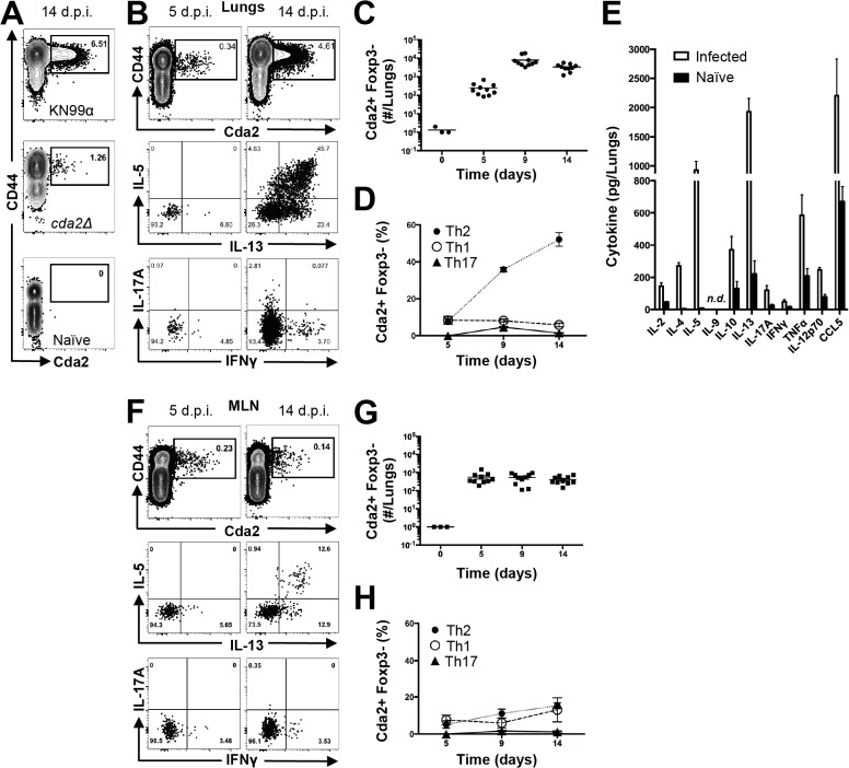
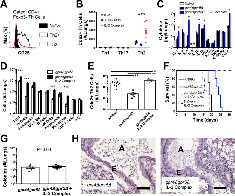
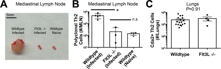
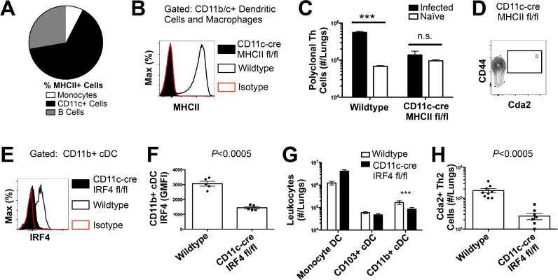
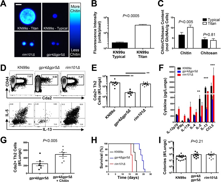
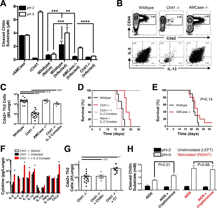
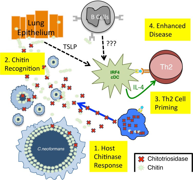
Pulmonary mycoses are often associated with type-2 helper T (Th2) cell responses. However, mechanisms of Th2 cell accumulation are multifactorial and incompletely known. To investigate Th2 cell responses to pulmonary fungal infection, we developed a peptide-MHCII tetramer to track antigen-specific CD4+ T cells produced in response to infection with the fungal pathogen Cryptococcus neoformans. We noted massive accruement of pathologic cryptococcal antigen-specific Th2 cells in the lungs following infection that was coordinated by lung-resident CD11b+ IRF4-dependent conventional dendritic cells. Other researchers have demonstrated that this dendritic cell subset is also capable of priming protective Th17 cell responses to another pulmonary fungal infection, Aspergillus fumigatus. Thus, higher order detection of specific features of fungal infection by these dendritic cells must direct Th2 cell lineage commitment. Since chitin-containing parasites commonly elicit Th2 responses, we hypothesized that recognition of fungal chitin is an important determinant of Th2 cell-mediated mycosis. Using C. neoformans mutants or purified chitin, we found that chitin abundance impacted Th2 cell accumulation and disease. Importantly, we determined Th2 cell induction depended on cleavage of chitin via the mammalian chitinase, chitotriosidase, an enzyme that was also prevalent in humans experiencing overt cryptococcosis. The data presented herein offers a new perspective on fungal disease susceptibility, whereby chitin recognition via chitotriosidase leads to the initiation of harmful Th2 cell differentiation by CD11b+ conventional dendritic cells in response to pulmonary fungal infection.


Conflict of interest statement

The authors have declared that no competing interests exist.

Figures

Fig 1
Fig 1. Type-2 Helper T Cells Accumulate in the Lungs of Mice Infected with C. neoformans.
(A) Cda2-MHCII tetramer identifies C. neoformans-specific helper T (Th) cells from mice 14 days post-infection with strain KN99α, cda2Δ, or age-matched, naïve mice. Flow cytometry plot (B) or graphs (C&D) from lung digests showing CD4+, Foxp3-, CD44+ Cda2+ Th cells expressing Th1 (IFNγ), Th2 (IL-5 & IL-13), or Th17 (IL-17A) cytokines. (E) Cytokines from lung homogenates 14 days post-infection with KN99α or age-matched, naïve mice. Flow cytometry plot (F) and graphs (G&H) from mediastinal lymph node suspensions of Th cells expressing Th1, Th2, or Th17 cytokines. Data are presented as mean +/- standard error with 2 independent experiments of at least 5 mice per group. Cda = Chitin deacetylase, CCL = chemokine ligand, IFN = interferon, IL = interleukin, MLN = mediastinal lymph node, TNF = tumor necrosis factor.
Fig 2
Fig 2. IL-2 Complexes Augment Type-2 Helper T Cells and Enhance Fungal Disease.
(A) CD25 expression by naïve (CD4+, CD44−), Th2- (CD4+, CD44+, IL-5-, IL-13−), and Th2+ (CD4+, CD44+, IL-5+, IL-13+) cells. (B) Cda+ Th cell numbers in the lungs of gpr4Δgpr5Δ infected mice treated with 5 μg IL-2, 25 μg anti-IL-2 antibody (JES6-1A12), or both (IL-2 Complex). Th1, Th17, and Th2 cells were identified by production of IFNγ, IL-17A, and IL-5/13, respectively. (C) Cytokines from lung homogenates of mice infected with gpr4Δgpr5Δ or age-matched naïve animals, with or without IL-2 complex treatment. (D) Pulmonary leukocytes from mice infected with gpr4Δgpr5Δ, with or without IL-2 complex treatment. (E) Cda2+ Th2 cells from lungs of mice infected with fully virulent KN99α, attenuated gpr4Δgpr5Δ, or mice infected with gpr4Δgpr5Δ and treated with IL-2 complex. (F) Survival of naïve mice or mice infected with gpr4Δgpr5Δ either with or without IL-2 complex treatment. P-value represents log-rank test comparing each survival curve with 10 mice per group to: gpr4Δgpr5Δ vs. gpr4Δgpr5Δ + IL-2 complex, P<0.0005; gpr4Δgpr5Δ vs. KN99α, P<0.0005; gpr4Δgpr5Δ + IL-2 complex vs. KN99α, P = 0.23. (G) Fungal burden within lungs of mice with or without IL-2 complex 14 days post-infection with gpr4Δgpr5Δ. (H) Hematoxylin and eosin staining of lungs from mice infected with gpr4Δgpr5Δ or similarly infected and treated with IL-2 complexes. A = brochial airway, E = epithelial cell layer. Data are presented as the mean +/- standard error with at least 2 independent experiments per group. * = P < 0.05, ** = P < 0.005, *** = P < 0.0005 by Mann-Whitney U. Cda = chitin deacetylase, CCL = chemokine ligand, DC = Dendritic Cells, IFN = interferon, IL = interleukin, ILC = Innate Lymphoid Cells, MΦ = Macrophages, NK = Natural Killer. TNF = tumor necrosis factor.
Fig 3
Fig 3. Lymphoid Priming is Dispensible for Pulmonary Th2 Cell Induction during Cryptococcal Infection.
(A) Lymphadenopathy of mediastinal lymph nodes (MLN) after 14 days post-infection with strain KN99α. (B) Quantification of IL-5+ IL-13+ polyclonal Th2 cells from the MLN in wild-type and Flt3L−/− mice. (C) Quantification of IL-5+ IL-13+ antigen-specific Th2 cells contained in the lungs of wild-type and Flt3L−/− mice.
Fig 4
Fig 4. Interferon Regulatory Factor 4-Dependent Conventional Dendritic Cells Coordinate Th2 Cell Induction.
(A) Antigen-presenting cells as a proportion of total MHCII+ cells in the lungs of infected mice. (B) MHCII expression by CD11b/c+ cells from mice 14 days post-infection with KN99α. Isotype refers to CD11b/c+ cells from wildtype mice stained with a rat IgG2b antibody of irrelevant specificity. (C) Quantification of pulmonary CD4+ Foxp3- CD44+ pulmonary Th cells. (D) Biexponential plot of CD4+ Foxp3- cells, indicating the absence of antigen-specific (CD44−, Cda2−) cells in the lungs of CD11c-cre MHCII mice infected with KN99α. (E) IRF4 expression by CD11b+ cDC from lungs of CD11cre IRF4 fl/fl or wildtype mice 14 days post-infection with KN99α. Isotype refers to CD11b+ cDC from wildtype mice stained with a rat IgG1 antibody of irrelevant specificity. (F) Geometric mean fluorescence intensity of CD11b+ cDC in from the lungs of infected CD11c-re IRF4 fl/fl mice. (G) Quantification of monocytes and dendritic cell subsets from the lungs of mutant or wildtype mice infected with KN99α. (H) Quantification of IL-5+ IL-13+ antigen-specific Th2 cells contained in the lungs of mutant or wildtype mice 14 days post-infection with KN99α. Data are presented as mean +/- standard error. * = P < 0.05, *** = P < 0.0005, and n.s. = not significant by Mann-Whitney U. Cda = chitin deacetylase, Flt3L = fms-like tyrosine kinase 3 ligand, MHC = major histocompatibility complex, cDC = conventional dendritic cells, IRF4 = interferon regulatory factor 4.
Fig 5
Fig 5. C. neoformans Chitin Correlates with Type-2 Helper T Cell Response and Subsequent Disease.
(A&B) Cryptococcal cells isolated from lungs at 14 days post-infection and stained with Calcofluor White. Cells were either imaged with epifluorescence microscopy (A) or analyzed by flow cytometry with fluorescence intensity calculated per pixel to determine chitin density within the cell wall (B). (C) Total chitin and chitosan content normalized to dry weight of cryptococccal cells. (D) Flow cytometry plots of CD4+, Foxp3-, CD44+ Cda2+ Th cells expressing Th2 cytokines, IL-5 & IL-13, at 14 days post-infection and (E) the quantification of these plots. (F) Cytokines from lung homogenates of mice 14 days post-infection. (G) IL-5+ IL-13+ antigen-specific Th2 cells from lungs of mice 14 days post-infection without and with intranasal chitin particle treatment. (H) P-value represents log-rank test comparing each survival curve with 10 mice per group to: gpr4Δgpr5Δ vs. KN99α, P<0.0005; gpr4Δgpr5Δ vs. rim101Δ, P<0.0005; KN99α vs. rim101Δ, P<0.0005. (I) Fungal burden in the lungs at 14 days post-infection. Data are presented as the mean +/- standard error with at least 2 independent experiments per group. ** = P < 0.005, *** = P < 0.0005 by Mann-Whitney U or Kruskal Wallis ANOVA. Cda = chitin deacetylase, CCL = chemokine ligand, GlcNAc = N-acetylglucosamine, IFN = interferon, IL = interleukin.
Fig 6
Fig 6. Chitotriosidase Promotes Chitin Recognition and Th2 Cell-mediated Disease.
(A) Chitinase enzyme activity of recombinant enzymes or lung homogenates from 14 days post-infected or naïve mice. (B) Flow cytometry plots of CD4+, Foxp3-, CD44+ Cda2+ Th cells expressing Th2 cytokines, IL-5 & IL-13, at 14 days post-infection and (C) the quantification of these plots. (D&E) Survival of mutant or wildtype mice infected with KN99α either with or without IL-2 complex treatment. Logrank test comparing each survival curve relative with 10 mice for each group to: Chit1 −/− vs. wildtype, P<0.005; Chit1 −/− vs. Chit1 −/− + IL-2 complex, P<0.0005; Chit1 −/− + IL-2 complex vs. wildtype, P = 0.07. (F) Cytokines from lung homogenates of naïve or wildtype and Chit1−/− mice 14 days post-infection with KN99α or age-matched, naïve Chit1−/− mice. (G) IL-5+ IL-13+ antigen-specific Th2 cells from lungs of Chit1−/− mice 14 days post-infection and mice treated with intranasal chitin particles or chitin heptamer fragments (C7). (H) Chitinase enzyme activity of human plasma without (LEFT) or with (RIGHT) cryptococcal lysate antigen stimulation. Data are presented as the mean +/- standard error with at least 2 independent experiments per group. * = P < 0.05, ** = P < 0.005, *** = P < 0.0005 by Mann-Whitney U or Kruskal Wallis ANOVA. AMCase = Acidic Mammalian Chitinase, C7 = chitin heptamer, CCL = chemokine ligand, Cda = chitin deacetylase, Chit1 = Chitotriosidase, IFN = interferon, IL = interleukin, TNF = tumor necrosis factor.
Fig 7
Fig 7. Model of Th2 Cell-mediated Disease during Pulmonary Fungal Infection.
1) Chitotriosidase is released by the host and degrades fungal chitin to generate small chitin fragments, such as chitin heptamers. 2) Chitin recognition occurs by an unknown mechanism that could involve either epithelial cell production of alarmins, such as thymic stromal lymphopoietin (TLSP) [9] or antibodies that bind chitin [9]. 3) These chitin-based signals are recognized by lung-resident CD11b+ conventional dendritic cells that are capable of producing IL-4, an essential Th2 cell differentiation factor. 4) The adaptive Th2 cell response results in enhanced disease in the absence of significant changes in pulmonary fungal burden.

References

    1. Brown GD, Denning DW, Gow NA, Levitz SM, Netea MG, et al. (2012) Hidden killers: human fungal infections. Sci Transl Med 4: 165rv113 10.1126/scitranslmed.3004404 - DOI - PubMed
    1. Denning DW, O'Driscoll BR, Hogaboam CM, Bowyer P, Niven RM (2006) The link between fungi and severe asthma: a summary of the evidence. Eur Respir J 27: 615–626. - PubMed
    1. Romani L (2011) Immunity to fungal infections. Nat Rev Immunol 11: 275–288. 10.1038/nri2939 - DOI - PubMed
    1. Licona-Limon P, Kim LK, Palm NW, Flavell RA (2013) TH2, allergy and group 2 innate lymphoid cells. Nat Immunol 14: 536–542. 10.1038/ni.2617 - DOI - PubMed
    1. Guilliams M, Ginhoux F, Jakubzick C, Naik SH, Onai N, et al. (2014) Dendritic cells, monocytes and macrophages: a unified nomenclature based on ontogeny. Nat Rev Immunol 14: 571–578. 10.1038/nri3712 - DOI - PMC - PubMed

Publication types

LinkOut - more resources