Pharmacological inhibition of ALDH1A in mice decreases all-trans retinoic acid concentrations in a tissue specific manner
- PMID: 25764981
- PMCID: PMC4420653
- DOI: 10.1016/j.bcp.2015.03.001
Pharmacological inhibition of ALDH1A in mice decreases all-trans retinoic acid concentrations in a tissue specific manner
Abstract
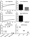
all-trans retinoic acid (atRA), the active metabolite of vitamin A, is an essential signaling molecule. Specifically the concentrations of atRA are spatiotemporally controlled in target tissues such as the liver and the testes. While the enzymes of the aldehyde dehydrogenase 1A family (ALDH1A) are believed to control the synthesis of atRA, a direct relationship between altered ALDH1A activity and tissue atRA concentrations has never been shown. To test whether inhibition of ALDH1A enzymes decreases atRA concentrations in a tissue specific manner, the potent ALDH1A inhibitor WIN 18,446 was used to inhibit ALDH1A activity in mice. The ALDH1A expression, atRA formation kinetics, ALDH1A inhibition by WIN 18,446 and WIN 18,446 disposition were used to predict the time course and extent of inhibition of atRA formation in the testis and liver. The effect of WIN 18,446 on atRA concentrations in testis, liver and serum were measured following single and multiple doses of WIN 18,446. ALDH1A1 and ALDH1A2 were responsible for the majority of atRA formation in the testis while ALDH1A1 and aldehyde oxidase contributed to atRA formation in the liver. Due to the different complement of enzymes contributing to atRA formation in different tissues and different inhibition of ALDH1A1 and ALDH1A2 by WIN 18,446, WIN 18,446 caused only a 50% decrease in liver atRA but testicular atRA decreased over 90%. Serum atRA concentrations were also reduced. These data demonstrate that inhibition of ALDH1A enzymes will decrease atRA concentrations in a tissue specific manner and selective ALDH1A inhibition could be used to alter atRA concentrations in select target tissues.
Keywords: Aldehyde dehydrogenase; Aldehyde oxidase; Liver; Mass spectrometry; Retinoic acid; Testes.
Copyright © 2015 Elsevier Inc. All rights reserved.
Figures


Similar articles
-
Aldehyde Oxidase Contributes to All-Trans-Retinoic Acid Biosynthesis in Human Liver.Drug Metab Dispos. 2021 Mar;49(3):202-211. doi: 10.1124/dmd.120.000296. Epub 2020 Dec 18. Drug Metab Dispos. 2021. PMID: 33355213 Free PMC article.
-
Sources of all-trans retinal oxidation independent of the aldehyde dehydrogenase 1A isozymes exist in the postnatal testis†.Biol Reprod. 2019 Feb 1;100(2):547-560. doi: 10.1093/biolre/ioy200. Biol Reprod. 2019. PMID: 30247516 Free PMC article.
-
Importance of ALDH1A enzymes in determining human testicular retinoic acid concentrations.J Lipid Res. 2015 Feb;56(2):342-57. doi: 10.1194/jlr.M054718. Epub 2014 Dec 11. J Lipid Res. 2015. PMID: 25502770 Free PMC article.
-
Role of Retinoic Acid-Metabolizing Cytochrome P450s, CYP26, in Inflammation and Cancer.Adv Pharmacol. 2015;74:373-412. doi: 10.1016/bs.apha.2015.04.006. Epub 2015 May 27. Adv Pharmacol. 2015. PMID: 26233912 Free PMC article. Review.
-
Aldehyde dehydrogenase 1A1: friend or foe to female metabolism?Nutrients. 2014 Mar 3;6(3):950-73. doi: 10.3390/nu6030950. Nutrients. 2014. PMID: 24594504 Free PMC article. Review.
Cited by
-
Design, synthesis, and ex vivo evaluation of a selective inhibitor for retinaldehyde dehydrogenase enzymes.Bioorg Med Chem. 2018 Dec 1;26(22):5766-5779. doi: 10.1016/j.bmc.2018.10.009. Epub 2018 Oct 24. Bioorg Med Chem. 2018. PMID: 30409702 Free PMC article.
-
Oncorequisite role of an aldehyde dehydrogenase in the pathogenesis of T-cell acute lymphoblastic leukemia.Haematologica. 2021 Jun 1;106(6):1545-1558. doi: 10.3324/haematol.2019.245639. Haematologica. 2021. PMID: 32414855 Free PMC article.
-
Aldehyde Oxidase Contributes to All-Trans-Retinoic Acid Biosynthesis in Human Liver.Drug Metab Dispos. 2021 Mar;49(3):202-211. doi: 10.1124/dmd.120.000296. Epub 2020 Dec 18. Drug Metab Dispos. 2021. PMID: 33355213 Free PMC article.
-
Evidence of depot-specific regulation of all-trans-retinoic acid biosynthesis in human adipose tissue.Clin Transl Sci. 2022 Jun;15(6):1460-1471. doi: 10.1111/cts.13259. Epub 2022 Mar 15. Clin Transl Sci. 2022. PMID: 35213790 Free PMC article.
-
Commensal segmented filamentous bacteria-derived retinoic acid primes host defense to intestinal infection.Cell Host Microbe. 2021 Dec 8;29(12):1744-1756.e5. doi: 10.1016/j.chom.2021.09.010. Epub 2021 Oct 21. Cell Host Microbe. 2021. PMID: 34678170 Free PMC article.
References
-
- Mucida D, Park Y, Kim G, Turovskaya O, Scott I, Kronenberg M, et al. Reciprocal TH17 and regulatory T cell differentiation mediated by retinoic acid. Science. 2007;317:256–260. - PubMed
-
- Iwata M, Hirakiyama A, Eshima Y, Kagechika H, Kato C, Song SY. Retinoic acid imprints gut-homing specificity on T cells. Immunity. 2004;21:527–538. - PubMed
Publication types
MeSH terms
Substances
Grants and funding
LinkOut - more resources
Full Text Sources
Other Literature Sources
Molecular Biology Databases
Miscellaneous

