. 2015 Apr 17;348(6232):347-52.
doi: 10.1126/science.aaa2958. Epub 2015 Mar 12.

Axonal regeneration. Systemic administration of epothilone B promotes axon regeneration after spinal cord injury

Affiliations

Axonal regeneration. Systemic administration of epothilone B promotes axon regeneration after spinal cord injury

Jörg Ruschel et al. Science. .

Abstract

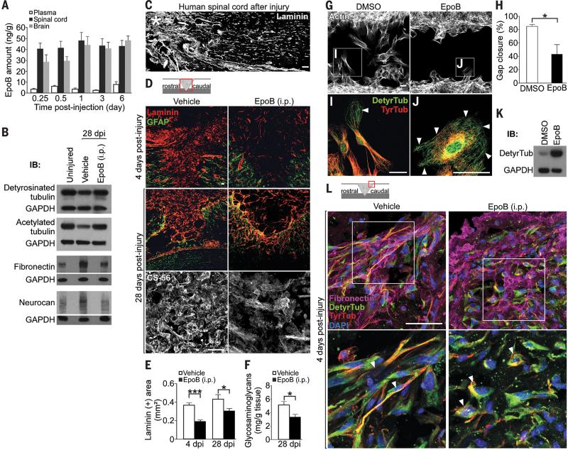
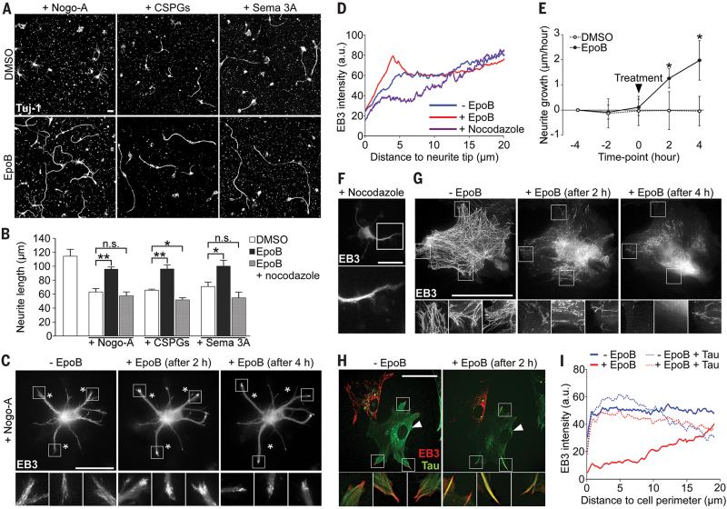
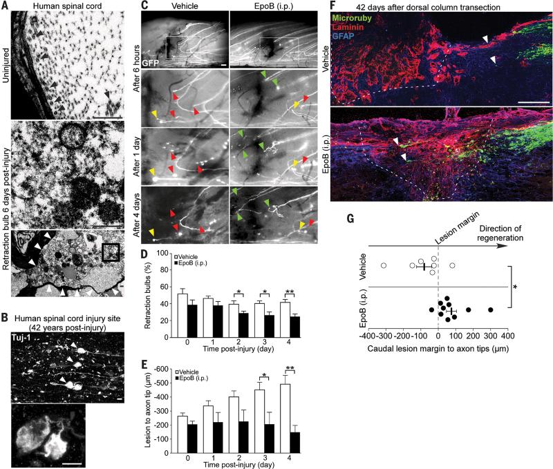
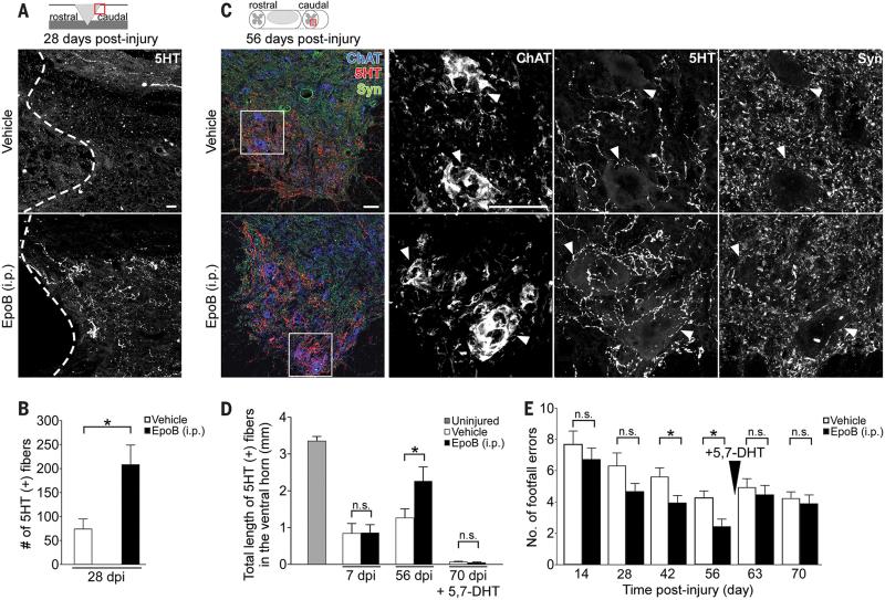
After central nervous system (CNS) injury, inhibitory factors in the lesion scar and poor axon growth potential prevent axon regeneration. Microtubule stabilization reduces scarring and promotes axon growth. However, the cellular mechanisms of this dual effect remain unclear. Here, delayed systemic administration of a blood-brain barrier-permeable microtubule-stabilizing drug, epothilone B (epoB), decreased scarring after rodent spinal cord injury (SCI) by abrogating polarization and directed migration of scar-forming fibroblasts. Conversely, epothilone B reactivated neuronal polarization by inducing concerted microtubule polymerization into the axon tip, which propelled axon growth through an inhibitory environment. Together, these drug-elicited effects promoted axon regeneration and improved motor function after SCI. With recent clinical approval, epothilones hold promise for clinical use after CNS injury.


Figures

Fig. 1
Fig. 1. Epothilone B increases microtubule stabilization, reduces fibrotic scar tissue and CSPG deposition after SCI by inhibiting meningeal fibroblast migration
(A) Mass spectrometric analysis of CNS tissue and blood plasma after a single epothilone B intraperitoneal injection, n = 4 animals per time-point. (B) Western blots (WB) of indicated proteins in pooled spinal cord (lesion) extracts, n = 3 animals per group. (C) Human spinal cord 42 years after injury (asterisk), laminin immunolabeling. (D) Immunolabeling for laminin, glial fibrillary acidic protein (GFAP) or chondroitin sulfates (CS-56) after rat spinal cord hemisection. (E) Laminin-immunopositive (+) area at the lesion site, n = 7 to 8 animals per group. (F) Glycosaminoglycan amounts in spinal cord lesion extracts, n = 8 animals per group. (G) Cultured meningeal fibroblasts migrating into a cell free area in wound healing assays. (H) Percentage of the area shown in (G) occupied with cells after 48 hours of migration, n = 3 experiments. (I and J) High magnification of the boxed areas in (G) showing tyrosinated (TyrTub) and detyrosinated tubulin (DetyrTub, white arrowheads). (K) Western blots of detyrosinated tubulin and GAPDH in dissociated meningeal fibroblasts 48 hours after indicated treatment. (L) Immunolabeling of fibronectin, detyrosinated and tyrosinated in the meninges at a rat spinal cord injury site. Bottom panel, high magnification of meningeal fibroblasts (arrowheads) in top panel. dpi, days post-injury. Schemes shown in (D) and (L) indicate lesion and displayed region (red box). Scale bars, 50 μm. Values are plotted as means ± SEM. *P < 0.05, ***P < 0.001.
Fig 2
Fig 2. Epothilone B promotes microtubule protrusion and axon elongation in neurons while dampening microtubule dynamics in scar-forming fibroblasts
(A) Beta-3 tubulin (Tuj-1) immunolabeling of neurons growing on inhibitory substrates (CSPGs, chondroitin sulfate proteoglycans; Sema 3A, Semaphorin 3A). (B) Neurite length of cortical neurons after 48 hours with (+) or without (-) indicated treatments, n = 3 to 4 experiments. (C) EB3-mCherry localization in a neuron cultured with Nogo-A, before and after epothilone B treatment. Bottom panels, high magnification of boxed areas in top panels. Asterisks, stable landmarks across time-points. (D) EB3-mCherry fluorescence intensity in neurites under indicated conditions, n = 9 to 16 neurons. (E) Neurite growth rates on Nogo-A before and after indicated treatment, n = 12 to 15 neurons. Black arrowhead, time of treatment. (F) EB3-mCherry localization in neurons treated with nocodazole. Bottom picture, high magnification of boxed area in top picture. (G and H) EB3- mCherry localization in cultured meningeal fibroblasts before and after epothilone B treatment. (G) Control cell, (H) control and tau-expressing (arrowhead) cells. Bottom panels, high magnification of boxed areas in top panels. (I) EB3-mCherry fluorescence intensity in fibroblast periphery under indicated conditions, n = 20 cells/condition. Scale bars, 25 μm. Values are plotted as means (+ SEM in (B) and (E)). *P < 0.05, **P < 0.01. n.s., not significant.
Fig. 3
Fig. 3. Epothilone B reduces dystrophy and promotes regeneration of injured spinal cord axons
(A) Electron microscope images of human spinal cord after injury. Top panel, undamaged axon containing microtubules (black arrowheads). Bottom panel, retraction bulb without microtubules (boxed area as high magnification in middle panel). Scale bars, 500 nm. (B) Beta-3 tubulin immunolabeling of retraction bulbs in chronic human SCI. Scale bar, 10 μm. (C) Lesioned GFP-positive spinal cord axons forming retraction bulbs (yellow arrowheads), dying back (red arrowheads) or regenerating (green arrowheads) over time. Boxed area in top panel outlines the displayed region of the panels below. Scale bars, 100 μm. (D and E) Percentage of injured axons forming retraction bulbs (D) and distance of the lesioned, proximal axon tip to the injury site (E), n = 8 animals per group. (F) Mouse spinal cord after injury, Microruby-traced dorsal column axons (white arrowheads), laminin and GFAP immunolabeling (dashed line, lesion border). Scale bar, 100 μm. (G) Average distance between the caudal lesion margin and the axonal tips in each animal, n = 7-10 animals per group. Values are plotted as means + SEM. *P < 0.05, **P < 0.01.
Fig. 4
Fig. 4. Epothilone B promotes regeneration of serotonergic spinal axons and enhances functional recovery of walking after spinal cord contusion
(A) Serotonin (5HT) immunolabeling (dashed line, lesion border) and (B) number of 5HT-labeled (+) fibers caudal to a rat spinal dorsal hemisection, n = 7-8 animals per group. (C) Coronal sections of the rat lumbar spinal cord after contusion injury. Left panel, coimmunostaining of 5HT, synaptophysin (Syn) and choline acetyltransferase (ChAT). Right panels, high magnification images of each marker in the boxed area (left panel) visualizing serotonergic innervation of motor neurons (arrowheads). (D) Total length of 5HT-immunopositive fibers in the spinal cord ventral horn after indicated treatments and time-points (5,7-DHT = 5,7-Dihydroxytryptamine), n = 4 (uninjured), 6 (7dpi) and 11-12 animals (56 and 70 dpi) per group, respectively. (E) Number of footfalls on the horizontal ladder, n = 10-11 animals per group. dpi, days post-injury. Schemes in (A) and (C) indicate lesion and displayed region (red box). Scale bars, 50 μm. Values are plotted as means + SEM. *P < 0.05. n.s., not significant.

Comment in

References

    1. Klapka N, et al. Suppression of fibrous scarring in spinal cord injury of rat promotes long-distance regeneration of corticospinal tract axons, rescue of primary motoneurons in somatosensory cortex and significant functional recovery. Eur J Neurosci. 2005 Dec;22:3047. - PubMed
    1. GrandPre T, Li S, Strittmatter SM. Nogo-66 receptor antagonist peptide promotes axonal regeneration. Nature. 2002;417:547. 05/30/print. - PubMed
    1. Bradbury EJ, et al. Chondroitinase ABC promotes functional recovery after spinal cord injury. Nature. 2002 Apr 11;416:636. - PubMed
    1. Schnell L, Schwab ME. Axonal regeneration in the rat spinal cord produced by an antibody against myelin-associated neurite growth inhibitors. Nature. 1990;343:269. 01/18/print. - PubMed
    1. Liu K, et al. PTEN deletion enhances the regenerative ability of adult corticospinal neurons. Nat Neurosci. 2010;13:1075. 09//print. - PMC - PubMed

Publication types

MeSH terms