. 2015 Feb 14;16(1):84.
doi: 10.1186/s12864-015-1262-5.

Induced pluripotent stem cell-derived neuronal cells from a sporadic Alzheimer's disease donor as a model for investigating AD-associated gene regulatory networks

Affiliations

Induced pluripotent stem cell-derived neuronal cells from a sporadic Alzheimer's disease donor as a model for investigating AD-associated gene regulatory networks

Amir M Hossini et al. BMC Genomics. .

Erratum in

Abstract

Background: Alzheimer's disease (AD) is a complex, irreversible neurodegenerative disorder. At present there are neither reliable markers to diagnose AD at an early stage nor therapy. To investigate underlying disease mechanisms, induced pluripotent stem cells (iPSCs) allow the generation of patient-derived neuronal cells in a dish.

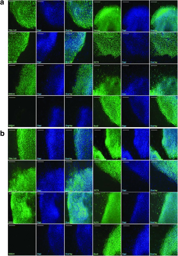
Results: In this study, employing iPS technology, we derived and characterized iPSCs from dermal fibroblasts of an 82-year-old female patient affected by sporadic AD. The AD-iPSCs were differentiated into neuronal cells, in order to generate disease-specific protein association networks modeling the molecular pathology on the transcriptome level of AD, to analyse the reflection of the disease phenotype in gene expression in AD-iPS neuronal cells, in particular in the ubiquitin-proteasome system (UPS), and to address expression of typical AD proteins. We detected the expression of p-tau and GSK3B, a physiological kinase of tau, in neuronal cells derived from AD-iPSCs. Treatment of neuronal cells differentiated from AD-iPSCs with an inhibitor of γ-secretase resulted in the down-regulation of p-tau. Transcriptome analysis of AD-iPS derived neuronal cells revealed significant changes in the expression of genes associated with AD and with the constitutive as well as the inducible subunits of the proteasome complex. The neuronal cells expressed numerous genes associated with sub-regions within the brain thus suggesting the usefulness of our in-vitro model. Moreover, an AD-related protein interaction network composed of APP and GSK3B among others could be generated using neuronal cells differentiated from two AD-iPS cell lines.

Conclusions: Our study demonstrates how an iPSC-based model system could represent (i) a tool to study the underlying molecular basis of sporadic AD, (ii) a platform for drug screening and toxicology studies which might unveil novel therapeutic avenues for this debilitating neuronal disorder.


Figures

Figure 1
Figure 1
Generation of human iPSCs from skin fibroblasts of a sporadic Alzheimer patient. (a): Morphology of fibroblasts NFH-46 in passage 4 (p4) before viral transduction. (b): Changes in morphology of NFH-46 seven days after infection with retroviruses. (c): Changes of NFH-46 on day 24 after infection shown in circle with arrow. (d): Typical image of non-embryonic stem cell like colony. (e, f): Typical morphology of AD-iPS colonies (AD-iPS-5, passage 4; AD-iPS-26B, passage 3) of one reprogramming experiment. (g): Typical morphology of AD-iPS colony in passage 3(p3). (h): AD-iPSC structure in high magnification. Scale bar, 100 μm.
Figure 2
Figure 2
AD-iPSCs express key pluripotency-associated proteins. Two AD-iPSC lines were successfully generated with one reprogramming experiment: AD-iPS5 (a) and AD-iPS26B (b). Both lines exhibited hESC-like morphologies, were positive for pluripotency-associated marker proteins, such as TRA-1-81, TRA-1-60, SSEA4, and NANOG, and were negative for the differentiation-specific marker SSEA1. Scale bar, 100 μm.
Figure 3
Figure 3
Neuronal differentiation of AD-iPSCs. Induction of neuronal cells by simultaneous treatment with inhibitors of transforming growth factor (TGF)-β receptor and MEK1/2 for 4 weeks. Neuronal cells derived from AD-iPSCs (a): AD-iPS5 neurons and (b): AD-iPS26B neurons showed the expression of neuronal markers, including PAX6, NESTIN and -β-TUBULIN III in a similar fashion as neuronal cells derived from the hESC line (c): H9. The neuronal differentiation was carried out once per AD-iPS cell line or H9 cell line. Scale bar, 100 μm.
Figure 4
Figure 4
Expression of neuronal marker genes and genes associated with Alzheimer-related brain regions in AD-iPS neurons. (a) Cluster analysis of neuronal marker genes in AD-iPS neurons and H9 neurons based on Euclidean distance of microarray-based gene expression values. (b) A number of genes of the heatmap data in (a) were confirmed by real- time PCR analysis. Bars indicate the RNA level normalized to β-ACTIN first and compared to gene expression of adult brain tissue (non-diseased, male, 21 years; Amsbio). The samples of AD-iPS neurons and H9 neurons consisted of cRNA generated from RNA isolated from a single well of a single neuronal differentiation respectively. In addition, to our data we analyzed AD brain RNA (male, 87 years, diagnosed AD; Amsbio). (c) Brain regions associated with genes from the AD KEGG pathway (but neither in Huntington disease nor in Parkinson disease) down-regulated in AD-iPS5 compared to H9 neurons. (d) Brain regions associated with genes from the AD KEGG pathway (but neither in Huntington nor in Parkinson disease) down-regulated in AD-iPS26B compared to H9 neurons.
Figure 5
Figure 5
Treatment of AD neuronal cells with the γ-secretase-inhibitor compound E (CE). Western blot analysis was used to monitor the expression of phosphorylated tau (p-tau), non-phosphorylated tau, GSK3α/β and phosphorylated GSK3β in AD-iPS5 neurons (INC5) and AD-iPS26B neurons (INC26B). The samples were derived from one well of a single neuronal differentiation. The parental fibroblast cells (NFH-46) were included as control in addition to the positive control (PC) for p-tau represented by lysate from neuroblastoma. β-ACTIN and GAPDH and coomassie blue staining were used to confirm similar protein loading across samples.
Figure 6
Figure 6
Expression of Alzheimer risk genes in AD-iPSC derived neurons. Cluster analysis of Alzheimer risk genes in experiments AD-iPS5 neurons, iPS26B neurons and H9 neurons of one neuronal differentiation experiment each. Up and down-regulated transcripts are depicted in red and green, respectively. RNA from AD-iPS5 neurons, AD-iPS26B neurons and H9 neurons was hybridized onto an Illumina human-8 BeadChip version 3. Alzheimer-associated genes known from genome wide association studies were filtered from the microarray experiments of AD-iPS5 neurons, AD-iPS26B neurons and H9 neurons. Illumina detection p-values were mapped to a binary scale (0 = not expressed if p-value > 0.05, 1 = expressed if p-value < = 0.05). These values were clustered via the R heatmap2 function using Euclidean distance as distance measure.
Figure 7
Figure 7
Gene expression associated with Alzheimer, Parkinson and Huntington disease in AD-iPS5 neurons. Venn diagram of down-regulated genes in the comparison AD-iPS5 vs. H9 neurons in KEGG pathways Alzheimer disease (AD), Parkinson disease (PD) and Huntington disease (HD). RNA from AD-iPS5 neurons and H9 neurons of one neuronal differentiation experiment was hybridized onto an Illumina human-8 BeadChip version 3. Functional annotation of significantly down-regulated genes from the experiments AD-iPS5 neurons vs. H9 neurons was performed with the DAVID Bioinformatics Resources 6.7. ips5 vs. H9: numbers of significant genes from KEGG pathways for ALzheimer, Parkinsons and Huntington.
Figure 8
Figure 8
Gene expression associated with Alzheimer, Parkinson and Huntington disease in AD-iPS26B neurons. Venn diagram of down-regulated genes in the comparison AD-iPS26B vs. H9 neurons in KEGG pathways Alzheimer disease (AD), Parkinson disease (PD) and Huntington disease (HD). RNA from AD-iPS26B neurons and H9 neurons was hybridized onto an Illumina human-8 BeadChip version 3. Functional annotation of significantly down-regulated genes from the experiments AD-iPS26B neurons vs. H9 neurons was performed with the DAVID Bioinformatics Resources 6.7. ips26 vs. H9: numbers of significant genes from KEGG pathways for ALzheimer, Parkinsons and Huntington.
Figure 9
Figure 9
Alzheimer-related protein association network in AD-iPS5 neurons. Protein association network retrieved from STRING v9 using genes from the Alzheimer disease pathway down-regulated in the AD-iPS5 neurons vs. H9 neurons comparison of one neuronal differentiation each. The network circles represent proteins. The lines between the circles show the functional association. Co-expression evidence: black, database evidence: light blue, textmining evidence: yellow, experimental evidence: purple, co-occurrence evidence: blue, neighborhood evidence: green, fusion evidence: red.
Figure 10
Figure 10
Alzheimer-related protein association network in AD-iPS26B neurons. Protein association network retrieved from STRING v9 using genes from the Alzheimer disease pathway down-regulated in the AD-iPS26B neurons vs. H9 neurons comparison of one neuronal differentiation each. The network circles represent proteins. The lines between the circles show the functional association. Co-expression evidence: black, database evidence: light blue, textmining evidence: yellow, experimental evidence: purple, co-occurrence evidence: blue, neighborhood evidence: green, fusion evidence: red.
Figure 11
Figure 11
Overlapping Alzheimer-related protein association network of AD-iPS5 neurons and of AD-iPS26B neurons. Protein association network of genes down-regulated in both AD-iPS5 neurons vs. H9 neurons and in AD-iPS26B neurons vs. H9 neurons comparisons of one neuronal differentiation. Protein interaction network retrieved from STRING v9 using genes from the Alzheimer disease pathway down-regulated in AD-iPS5 vs. H9 neurons and in AD-iPS26B vs. H9 neurons experiments. The network circles represent proteins. The lines between the circles show the functional association. Co-expression evidence: black, database evidence: light blue, textmining evidence: yellow, experimental evidence: purple, co-occurrence evidence: blue, neighborhood evidence: green, fusion evidence: red.
Figure 12
Figure 12
UPS-associated gene expression in AD-iPSC neurons. Cluster analysis based on Pearson correlation of UPS-related genes in AD-iPS neurons and H9 neurons. Up and down-regulated transcripts are depicted in red and green, respectively. RNA from AD-iPS5 neurons, AD-iPS26B neurons and H9 neurons of one well of one neuronal differentiation each was hybridized onto an Illumina human-8 BeadChip version 3. Known proteasome-related genes were selected from the microarray experiments of AD-iPS5 neurons, AD-iPS26B neurons and H9 neurons. Their Illumina average signal intensities were transformed to a logarithmic scale (log2) and clustered with the R heatmap2 function using Pearson correlation as similarity measure.

References

    1. Blennow K, de Leon MJ, Zetterberg H. Alzheimer’s disease. Lancet. 2006;368(9533):387–403. doi: 10.1016/S0140-6736(06)69113-7. - DOI - PubMed
    1. Selkoe DJ. Alzheimer’s disease results from the cerebral accumulation and cytotoxicity of amyloid beta-protein. J Alzheimers Dis. 2001;3(1):75–80. - PubMed
    1. Gatz M, Reynolds CA, Fratiglioni L, Johansson B, Mortimer JA, Berg S, et al. Role of genes and environments for explaining Alzheimer disease. Arch Gen Psychiatry. 2006;63(2):168–74. doi: 10.1001/archpsyc.63.2.168. - DOI - PubMed
    1. Cruts M, van Duijn CM, Backhovens H, Van den Broeck M, Wehnert A, Serneels S, et al. Estimation of the genetic contribution of presenilin-1 and −2 mutations in a population-based study of presenile Alzheimer disease. Hum Mol Genet. 1998;7(1):43–51. doi: 10.1093/hmg/7.1.43. - DOI - PubMed
    1. Selkoe DJ. Alzheimer’s disease: genes, proteins, and therapy. Physiol Rev. 2001;81(2):741–66. - PubMed

Publication types

MeSH terms

Substances