. 2015 May;138(Pt 5):1284-96.
doi: 10.1093/brain/awv047. Epub 2015 Mar 12.

The integrative role of the pedunculopontine nucleus in human gait

Affiliations

The integrative role of the pedunculopontine nucleus in human gait

Brian Lau et al. Brain. 2015 May.

Abstract

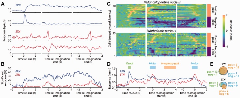
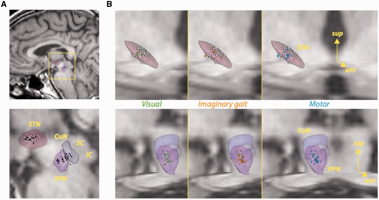
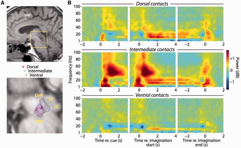
The brainstem pedunculopontine nucleus has a likely, although unclear, role in gait control, and is a potential deep brain stimulation target for treating resistant gait disorders. These disorders are a major therapeutic challenge for the ageing population, especially in Parkinson's disease where gait and balance disorders can become resistant to both dopaminergic medication and subthalamic nucleus stimulation. Here, we present electrophysiological evidence that the pedunculopontine and subthalamic nuclei are involved in distinct aspects of gait using a locomotor imagery task in 14 patients with Parkinson's disease undergoing surgery for the implantation of pedunculopontine or subthalamic nuclei deep brain stimulation electrodes. We performed electrophysiological recordings in two phases, once during surgery, and again several days after surgery in a subset of patients. The majority of pedunculopontine nucleus neurons (57%) recorded intrasurgically exhibited changes in activity related to different task components, with 29% modulated during visual stimulation, 41% modulated during voluntary hand movement, and 49% modulated during imaginary gait. Pedunculopontine nucleus local field potentials recorded post-surgically were modulated in the beta and gamma bands during visual and motor events, and we observed alpha and beta band synchronization that was sustained for the duration of imaginary gait and spatially localized within the pedunculopontine nucleus. In contrast, significantly fewer subthalamic nucleus neurons (27%) recorded intrasurgically were modulated during the locomotor imagery, with most increasing or decreasing activity phasically during the hand movement that initiated or terminated imaginary gait. Our data support the hypothesis that the pedunculopontine nucleus influences gait control in manners extending beyond simply driving pattern generation. In contrast, the subthalamic nucleus seems to control movement execution that is not likely to be gait-specific. These data highlight the crucial role of these two nuclei in motor control and shed light on the complex functions of the lateral mesencephalus in humans.

Keywords: Parkinson’s disease; deep brain stimulation; gait; pedunculopontine nucleus; subthalamic nucleus.


Figures

None
Gait disorders are untreatable in the elderly because the physiology of gait control is poorly understood. Lau et al. present electrophysiological evidence that the brainstem pedunculopontine nucleus is modulated by visual stimulation, voluntary movement and imaginary gait, suggesting that the brainstem controls gait in manners extending beyond driving pattern generation.
Figure 1
Figure 1
Neural responses in the human PPN and STN during imaginary gait. (A) Example PPN and STN activity. Each row represents a different neuron, with each column illustrating activity aligned to one of three different trial events. Activity was smoothed with an optimal Gaussian kernel and averaged over trials. We pooled data from normal and rapid gait imagination since we did not observe any differences between these conditions. (B) The PPN is more responsive than the STN during the imaginary gait task. For each neuron, we compared activity at each point in time with the baseline activity of that neuron taken before the presentation of the instruction cue. Significance was pooled across all neural responses after FDR correction, and smoothed with a 50 ms moving average. (C) Individual activity profiles for significantly modulated neural responses. All neurons that were significantly modulated relative to baseline are plotted, aligning to the same events as A and B. The activity for each neural response is smoothed with an optimal Gaussian kernel and normalized to the peak absolute response across the entire trial. In each panel, the neural responses are sorted according to the time of the peak absolute response (separately for positive and negative peak responses). The sign of the peak absolute response is indicated to the right of the last column. (D) The PPN and STN respond differently during imaginary gait. Population activity profile including all neurons that were significantly modulated relative to baseline. Neural activity was z-scored relative to baseline before averaging the absolute response. (E) Neural responses in the PPN more frequently occur to different task epochs than those in the STN. Individual neural responses were tested for significant differences during three different epochs (indicated in D): (i) Visual, corresponding to the presentation of the instruction cue (green); (ii) Motor, corresponding to the overt button presses that indicated the beginning and end of imaginary gait (blue); and (iii) Imaginary gait, when patients had their eyes closed and were imagining walking (orange). ‘Pos’ and ‘neg’ indicate the number of neurons exhibiting positive or negative modulations of activity for each epoch.
Figure 2
Figure 2
Localization of neurons in the STN and PPN. (A) Sagittal views of task-related (black) and non-task-related (white) neurons. (B) Localization within the STN (top row) or cuneiform nucleus (CuN)/PPN (bottom row) of neurons with significant modulations in activity during visual instruction (first column, green spheres), imaginary gait (second column, orange spheres), and button press (third column, blue spheres). White spheres represent the remaining neurons. The STN and CuN/PPN figures include axial and coronal T1-weighted MRI sections of the histological atlas of Yelnik et al. (2007). IC = inferior colliculus; SC = superior colliculus.
Figure 3
Figure 3
Task-related local field potential activity is spatially localized within the PPN. (A) Sagittal view of post-surgical localization of PPN electrodes in T1-weighted MRI image from one patient (top). Illustration of dipole centres for all patients in the histological atlas of Yelnik et al. (2007) (bottom). Each sphere represents the midway point between adjacent contacts contributing to each bipolar recording. (B) Spectrograms of local field potentials recorded from definitive DBS contacts. Recordings from each patient were averaged by dorsal-ventral arrangement along the electrode shaft (rows). The panels within each row are aligned to presentation of the visual instruction cue, initiation and termination of imaginary gait. Spectral power is normalized relative to prestimulus power (−3 to −1 s before instruction onset). IC = inferior colliculus; SC = superior colliculus.

Comment in

Similar articles

Cited by

References

    1. Androulidakis AG, Mazzone P, Litvak V, Penny W, Dileone M, Gaynor LMFD, et al. Oscillatory activity in the pedunculopontine area of patients with Parkinson’s disease. Exp Neurol. 2008;211:59–66. - PubMed
    1. Aravamuthan BR, Angelaki DE. Vestibular responses in the macaque pedunculopontine nucleus and central mesencephalic reticular formation. Neuroscience. 2012;223:183–99. - PMC - PubMed
    1. Aron AR, Poldrack RA. Cortical and subcortical contributions to Stop signal response inhibition: role of the subthalamic nucleus. J Neurosci. 2006;26:2424–33. - PMC - PubMed
    1. Bakker M, de Lange FP, Stevens JA, Toni I, Bloem BR. Motor imagery of gait: a quantitative approach. Exp Brain Res. 2007;179:497–504. - PubMed
    1. Bardinet E, Bhattacharjee M, Dormont D, Pidoux B, Malandain G, Schüpbach M, et al. A three-dimensional histological atlas of the human basal ganglia. II. Atlas deformation strategy and evaluation in deep brain stimulation for Parkinson disease. J Neurosurg. 2009;110:208–19. - PubMed

Publication types