. 2015 Mar 13:6:6463.
doi: 10.1038/ncomms7463.

Sensitive β-galactosidase-targeting fluorescence probe for visualizing small peritoneal metastatic tumours in vivo

Affiliations

Sensitive β-galactosidase-targeting fluorescence probe for visualizing small peritoneal metastatic tumours in vivo

Daisuke Asanuma et al. Nat Commun. .

Abstract

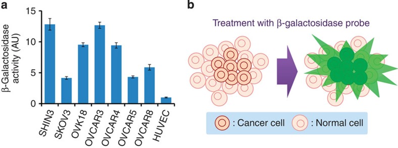
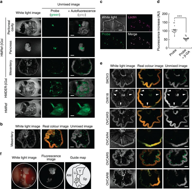
Fluorescence-guided diagnostics is one of the most promising approaches for facile detection of cancer in situ. Here we focus on β-galactosidase, which is overexpressed in primary ovarian cancers, as a molecular target for visualizing peritoneal metastases from ovarian cancers. As existing fluorescence probes are unsuitable, we have designed membrane-permeable HMRef-βGal, in which the optimized intramolecular spirocyclic function affords >1,400-fold fluorescence enhancement on activation. We confirm that HMRef-βGal sensitively detects intracellular β-galactosidase activity in several ovarian cancer lines. In vivo, this probe visualizes metastases as small as <1 mm in diameter in seven mouse models of disseminated human peritoneal ovarian cancer (SHIN3, SKOV3, OVK18, OVCAR3, OVCAR4, OVCAR5 and OVCAR8). Because of its high brightness, real-time detection of metastases with the naked eye is possible. Endoscopic fluorescence detection of metastases is also demonstrated. The results clearly indicate preclinical potential value of the probe for fluorescence-guided diagnosis of peritoneal metastases from ovarian cancers.


Figures

Figure 1
Figure 1. β-Galactosidase-targeting cancer visualization strategy.
(a) β-Galactosidase activity per protein abundance in lysate of SHIN3, SKOV3, OVK18, OVCAR3, OVCAR4, OVCAR5, OVCAR8 or HUVEC cells. Data represent mean±s.d. from a single experiment in triplicate. (b) Schematic illustration of fluorescence detection of cancer cells with enhanced β-galactosidase activity using a fluorescence probe.
Figure 2
Figure 2. Detection of β-galactosidase activities in living ovarian cancer cells with HMRef-βGal.
(a) Activation of HMRef-βGal on enzymatic reaction with β-galactosidase. Photographs show cuvettes containing the reaction mixture irradiated with 365-nm ultraviolet light. (b) Time course of enzymatic reaction of HMRef-βGal with β-galactosidase. β-Galactosidase (5 units) was added at 50 s. HMDER-βGal was used as a control. Probe concentrations were 0.5 μM. (c) Confocal images of ovarian tumour cells treated with HMRef-βGal. Cells were incubated with 10 μM HMRef-βGal for 1 h, and DIC and fluorescence images were obtained. Ex/Em=498 nm/505–600 nm. Scale bar, 100 μm. (d) Fluorescence intensity of ovarian cancer cells treated with HMRef-βGal in the presence or absence of β-GA. Fluorescence intensity of cells on a 96-well plate was measured with a plate reader (Ex/Em=498 nm/518 nm). Probe concentration was 10 μM. Data represent means±s.e.m (n=4).
Figure 3
Figure 3. Visualization of peritoneal metastases in mouse models with HMRef-βGal.
(a,b) Fluorescence spectral imaging of peritoneal SHIN3 metastasis with various probes. At 5 min (a) or 1 h post administration of probe (b), mice were immediately killed and imaged. In spectral unmixed images, fluorescence from the probe and autofluorescence were assigned as green and grey, respectively. Scale bar, 5 mm. (c) Dual-colour fluorescence imaging of metastases. Lectin-targeted staining was performed as a marker for metastases (Lectin). The mouse model was also treated for 1 h with HMRef-βGal and imaged (Probe). The merged image was prepared by overlaying the two unmixed images. Scale bar, 5 mm. (d) Fluorescence intensity on tumour nodules. In live mouse models, tumour nodules were treated with 100 μM HMRef-βGal in the presence or absence of 10 mM β-GA (see Supplementary Fig. 6). ***P<0.001 by Welch’s t-test (n=20 for each group). (e) Fluorescence spectral imaging of several mouse models of peritoneal metastasis at 1 h post administration of HMRef-βGal. Arrowheads indicate metastases in the OVK18 images. Scale bar, 5 mm. (f) Fluorescence endoscopy of tumours inside the peritoneal cavity. AW, abdominal wall; Li, liver; Pc, pancreas; Sp, spleen; T, tumour. See Supplementary Video S1 online for real-time monitoring endoscopy.

References

    1. Carmignani C. P., Sugarbaker T. A., Bromley C. M. & Sugarbaker P. H. Intraperitoneal cancer dissemination: mechanisms of the patterns of spread. Cancer Metastasis Rev. 22, 465–472 (2003) . - PubMed
    1. Cotte E., Passot G., Gilly F. N. & Glehen O. Selection of patients and staging of peritoneal surface malignancies. World J. Gastrointest. Oncol. 2, 31–35 (2010) . - PMC - PubMed
    1. Lengyel E. Ovarian cancer development and metastasis. Am. J. Pathol. 177, 1053–1064 (2010) . - PMC - PubMed
    1. Pecorelli S., Benedet J. L., Creasman W. T., Shephard J. H. & Pettersson F. Annual Report on The Results of Treatment in Gynecological Cancer Vol. 23, International Federation of Gynecology and Obstetrics (1998) .
    1. Halkia E., Spiliotis J. & Sugarbaker P. Diagnosis and management of peritoneal metastases from ovarian cancer. Gastroenterol. Res. Pract. 2012, 541842 (2012) . - PMC - PubMed

Publication types