Review
. 2015 Feb:30:134-146.
doi: 10.1016/j.sbi.2015.02.011. Epub 2015 Mar 10.

Characterizing excited conformational states of RNA by NMR spectroscopy

Affiliations
Review

Characterizing excited conformational states of RNA by NMR spectroscopy

Bo Zhao et al. Curr Opin Struct Biol. 2015 Feb.

Abstract

Conformational dynamics is a hallmark of diverse non-coding RNA functions. During these functional processes, RNA molecules almost ubiquitously undergo conformational transitions that are tuned to meet distinct structural and kinetic requirements for proper function. A complete mechanistic understanding of RNA function requires comprehensive structural and dynamic knowledge of these complex transitions, which often involve alternative higher-energy conformational states that pose a major challenge for high-resolution structural study by conventional methods. In this review, we describe recent progress in RNA NMR that has started to unveil detailed structural, thermodynamic and kinetic insights into some of these excited conformational states of RNA and their functional roles in biology.


Conflict of interest statement

Conflicts of interest

The authors declare no conflicts of interest.

Figures

Figure 1
Figure 1
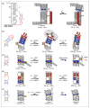
NMR characterization of excited conformational states in RNA. (a) A hierarchal free-energy landscape of RNA conformational transitions. Top panel: Tier-0 conformational transitions consist of large-scale changes in secondary structures such as pseudoknot formation from a hairpin. Due to high-energy barriers separating different secondary structures, conformational transition from a Tier-0 ground state (GS) to a Tier-0 excited state (ES) occurs at the timescale of tens-of-milliseconds to seconds and longer. Middle panel: Tier-1 conformational transitions consist of local base-pairing re-configurations such as base pair reshuffling within a hairpin. The energy barriers between Tier-1 states are smaller than those between Tier-0 states, and the resulting conformational transition from a Tier-1 GS to a Tier-1 ES occurs at the timescale of microseconds to tens-of-milliseconds. Bottom panel: Tier-2 conformational transitions consist of fine-tuned conformational changes such as base stacking dynamics and interhelical motions. With energy barriers of only a few kBT, conformational transition from a Tier-2 GS to a Tier-2 ES occurs at the timescale of picoseconds to microseconds. (b) Solution-state NMR techniques for characterizing excited conformational states in RNA. Left panel: timescales of exchange processes and corresponding suitable NMR experiments (solid lines). Dashed lines indicate regions that are challenging to study by a given NMR experiment. Right panel: schematic representation of experimental profiles measured with different NMR techniques for characterizing excited states in RNA. NMR resonance for a sparsely populated and shortly lived excited state, which is not visible in a conventional Heteronuclear Single Quantum Coherence (HSQC) spectrum, is illustrated as red dashed circles for clarity.
Figure 2
Figure 2
NMR characterization of Tier-0 excited conformational states. (a) The Vibrio vulnificus add adenine riboswitch in the absence of ligand undergoes a temperature-dependent conformational exchange between a ground state and a minor state at a rate of 1.31 s−1 at 25 °C. The ground state is a binding-inactive translational-off state, and has a population ranging between ~90% at 10 °C to ~60% at 40 °C. The minor or excited state is a binding-active translational-off state, and has a population ranging between ~10% at 10 °C to ~40% at 40 °C. The minor/excited state is subsequently switched to the translational-on state in the presence of adenine. (b) The Fusobacterium nucleatum preQ1 riboswitch aptamer domain in the absence of ligand undergoes a conformational exchange between an unfolded ground state (pG ~ 90%) and a pseudoknot excited state (pE ~ 0%) at 25 °C. A site-specific labeling strategy was applied on residues A3, A10, U20, and U32 to facilitate secondary structure confirmation of a proposed pseudoknot excited state of the ligand-free aptamer. As shown in dashed arrows, the pseudoknot state may be selected by preQ1 during adaptive recognition toward the ligand-bound active state. (c) The Bacillus cereus fluoride riboswitch aptamer in the absence of ligand undergoes a conformational exchange between an unfolded ground state ( pG = 90%) and a potential pseudoknot-like excited state (pE = 10%) at a rate of 112 s−1 at 30 °C. This excited state is NMR ‘invisible’, and its detection and identification were obtained based on 13C CEST NMR spectroscopy. As shown in dashed arrows, this potential pseudoknot excited state may also be selected by fluoride and magnesium during adaptive recognition toward the ligand-bound active state.
Figure 3
Figure 3
NMR characterization of high-tier excited conformational states. (a–d) Tier-1 excited conformational states. (a) The MLV recoding signal has an overall pseudoknot secondary structure that undergoes a pH-dependent conformational exchange between a ground state ( pG ~ 94%) and an excited state (pE ~ 6%) at physiological pH. The ground state has a loose pseudoknot conformation that inactivates the readthrough by the ribosome to produce only Gag protein. The excited state has a compact pseudoknot conformation with a base triple between protonated A17 and residues C23 and G53 as well as tertiary interactions between L2 loop and S1 helix. The compact pseudoknot excited state interacts with the ribosome to ensure the later to bypass the stop codon for producing Gag-Pol fusion protein. (b) The HIV-1 TAR RNA has an overall hairpin secondary structure that undergoes complex conformational exchanges between one ground state (pG = 86.6%) and two excited states (pE1 = 13% and pE2 = 0.4%). The ground state has a hexanucleotide loop that allows key residues C30, U31, G34, and A35 to interact with Tat and Cyclin T1. The apical loop of HIV-1 TAR undergoes a microsecond-timescale exchange toward the first excited state (ES1), which sequesters key loop residues to form an autoinhibited state that prevents transactivation of the HIV-1 genome. The two bulge residues and the entire upper helix undergoes a millisecond-timescale exchange toward the second excited state (ES2), which has a global register shift with the formation of four non-canonical base pairs. (c) The ribosomal A-site internal loop undergoes a conformational exchange between a ground state (pG = 97.5%) and an excited state ( pE = 2.5%). The ground state has a flexible bulge of A1492 (A92) and A1493 (A93) that can flip out to interact with the codon–anticodon base pair between tRNA and mRNA for translational fidelity. The excited state flips out residue U95 and sequesteres both A92 and A93 into a helical conformation, which impairs the decoding and disrupts formation of the B2a intersubunit bridge as highlighted by the green dashed line. (d) The HIV-1 SL1 undergoes complex conformational exchange between one ground state (pG = 89%) and two excited states (pE1 = 9% and pE2 = 2%) that are centered on seven base pairs around the SL1 bulge. While the GS has a central-located internal bulge, ES1 and ES2 have upward and downward migrated bulges, respectively. This branched three-state reshuffling process generates a ‘moving zipper’ that provides a mechanism for ATP-independent HIV-1 SL1 isomerization. (e) A Tier-2 excited conformational state in the U6 spliceosomal RNA ISL. The U6 SL1 undergoes a conformational exchange between the ground state and the excited state that features a looped out residue U80 and a protonated residue A79 that forms a non-canonical base pair with C67. The looping out process of residue U80 may provide a pH-sensitive switch for controlling Mg2+ binding at the U6 ISL RNA.

Similar articles

Cited by

References

    1. Kruger K, Grabowski PJ, Zaug AJ, Sands J, Gottschling DE, Cech TR. Self-splicing RNA: autoexcision and autocyclization of the ribosomal RNA intervening sequence of Tetrahymena. Cell. 1982;31:147–157. - PubMed
    1. Guerrier-Takada C, Gardiner K, Marsh T, Pace N, Altman S. The RNA moiety of ribonuclease P is the catalytic subunit of the enzyme. Cell. 1983;35:849–857. - PubMed
    1. Mattick JS. RNA regulation: a new genetics? Nat Rev Genet. 2004;5:316–323. - PubMed
    1. Sharp PA. The centrality of RNA. Cell. 2009;136:577–580. - PubMed
    1. Williamson JR. Induced fit in RNA–protein recognition. Nat Struct Biol. 2000;7:834–837. - PubMed

Publication types

Substances

LinkOut - more resources