Review
. 2015 Mar 13;47(3):e147.
doi: 10.1038/emm.2014.117.

Degradation of misfolded proteins in neurodegenerative diseases: therapeutic targets and strategies

Affiliations
Review

Degradation of misfolded proteins in neurodegenerative diseases: therapeutic targets and strategies

Aaron Ciechanover et al. Exp Mol Med. .

Abstract

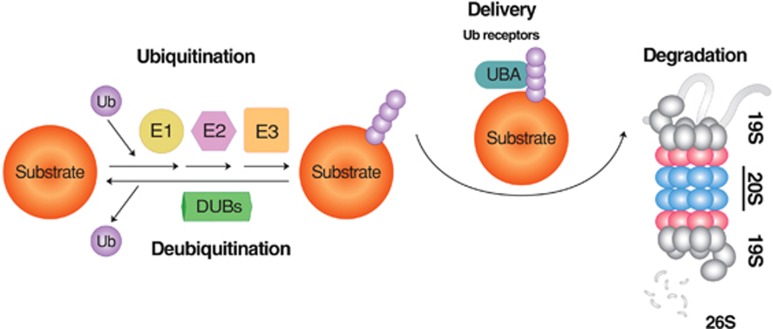
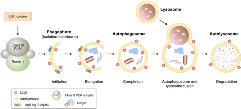
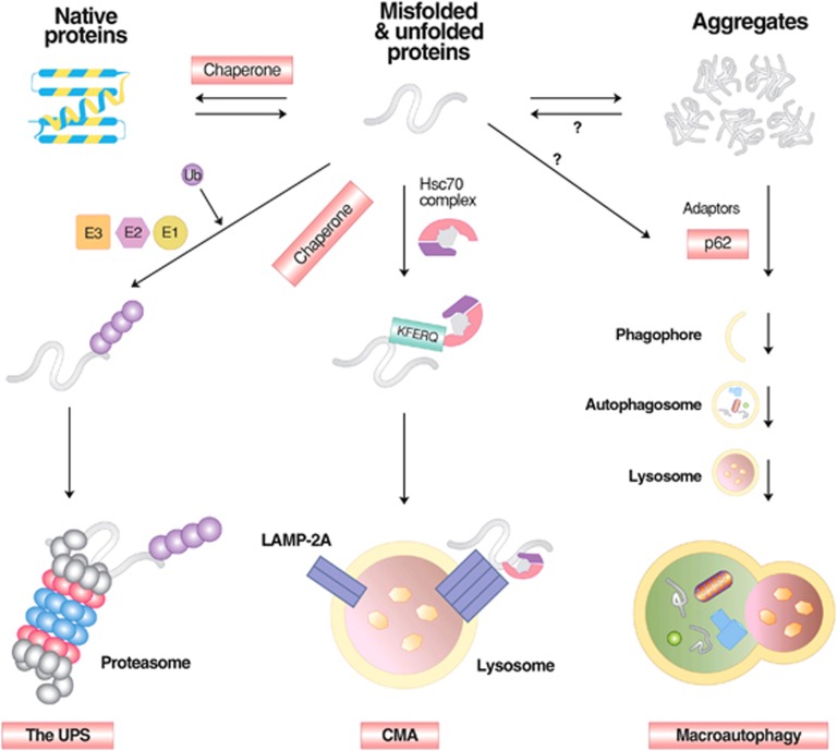
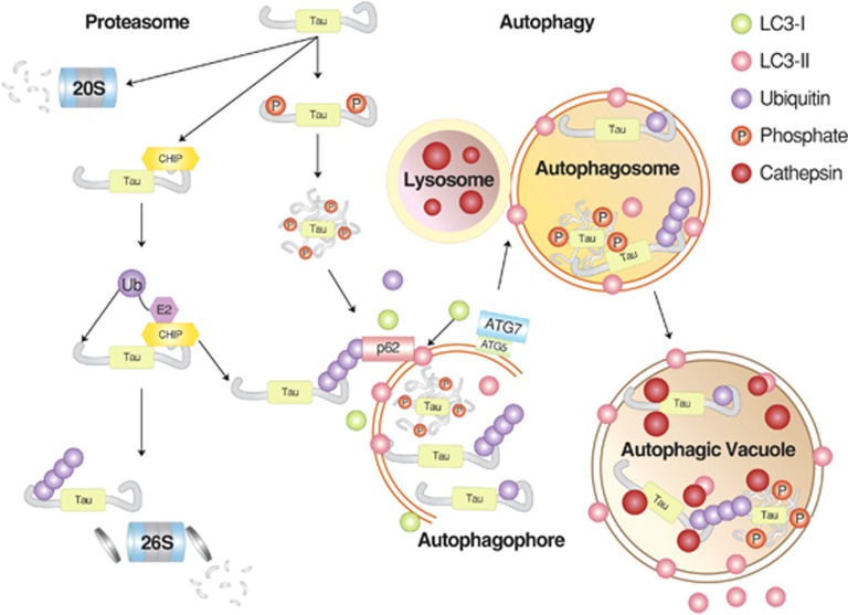
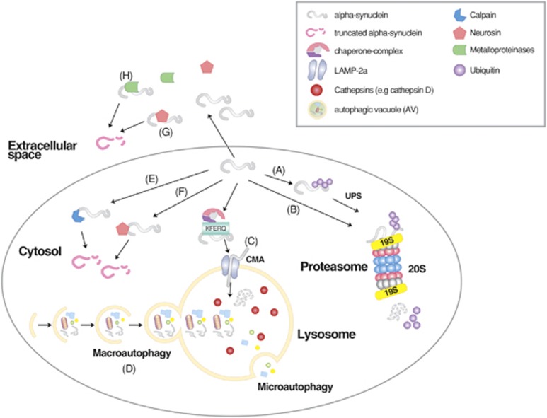
Mammalian cells remove misfolded proteins using various proteolytic systems, including the ubiquitin (Ub)-proteasome system (UPS), chaperone mediated autophagy (CMA) and macroautophagy. The majority of misfolded proteins are degraded by the UPS, in which Ub-conjugated substrates are deubiquitinated, unfolded and cleaved into small peptides when passing through the narrow chamber of the proteasome. The substrates that expose a specific degradation signal, the KFERQ sequence motif, can be delivered to and degraded in lysosomes via the CMA. Aggregation-prone substrates resistant to both the UPS and the CMA can be degraded by macroautophagy, in which cargoes are segregated into autophagosomes before degradation by lysosomal hydrolases. Although most misfolded and aggregated proteins in the human proteome can be degraded by cellular protein quality control, some native and mutant proteins prone to aggregation into β-sheet-enriched oligomers are resistant to all known proteolytic pathways and can thus grow into inclusion bodies or extracellular plaques. The accumulation of protease-resistant misfolded and aggregated proteins is a common mechanism underlying protein misfolding disorders, including neurodegenerative diseases such as Huntington's disease (HD), Alzheimer's disease (AD), Parkinson's disease (PD), prion diseases and Amyotrophic Lateral Sclerosis (ALS). In this review, we provide an overview of the proteolytic pathways in neurons, with an emphasis on the UPS, CMA and macroautophagy, and discuss the role of protein quality control in the degradation of pathogenic proteins in neurodegenerative diseases. Additionally, we examine existing putative therapeutic strategies to efficiently remove cytotoxic proteins from degenerating neurons.


Figures

Figure 1
Figure 1
The degradation of short-lived proteins by the UPS. In this selective proteolytic system, Ub is first activated by E1 and subsequently transferred to E2. In parallel, misfolded substrates of the UPS are recognized by molecular chaperones, such as CHIP, and associated with Ub ligases that promote the transfer of E2-conjugated Ub to specific Lys residues of substrates. Ubiquitinated substrates are deubiquitinated, unfolded, fed into the narrow chamber of the proteasome, and progressively cleaved into small peptides. Depending on the types of E3 ligases, Ub can be directly transferred from E2 to the substrate or via a two-step process that involves a transient binding of E3 to Ub. The repetition of this reaction results in the growth of a singly conjugated Ub to a chain of Ub with different topologies, depending on how Ub is conjugated to another Ub. Modified from Wang and Robbins.
Figure 2
Figure 2
Autophagosome formation and lysosomal degradation. Autophagosome formation can be triggered when the mTOR complex is inhibited by various stressors, such as starvation. This induces the assembly of the ULK protein complex composed of ULK1, Atg13 and FIP200 at the isolation membrane, which, in turn, activates the formation of the Beclin-1/PI3KC3 complex composed of Beclin-1, UVRAG, Bif-1, Ambra1, Vps15 and Vps34. During the elongation of the isolation membrane, the Atg5-Atg12-Atg16L1 complex mediates the conjugation of PE to LC3-I, generating LC3-II that relocates from the cytosol to the autophagic membrane and is anchored on its surface. The resulting autophagic membrane structures—autophagosomes—are fused with lysosomes to form autolysosomes, wherein cargoes, including misfolded proteins, are degraded by lysosomal hydrolases.
Figure 3
Figure 3
The degradation of misfolded proteins by various cellular proteolytic pathways. Misfolded proteins are initially recognized by molecular chaperones that deliver the substrates to the UPS, CMA or macroautophagy depending on the nature of misfolding, size and solubility. In general, soluble and monomeric misfolded proteins are primarily degraded by the UPS and CMA. In CMA, substrates carrying the KFERQ motif are recognized and bound by Hsc70 in association with chaperones. The substrates are subsequently delivered to the LAMP2 complex on the lysosomal membrane, translocated to the lumen, and degraded into amino acids by lysosomal hydrolases. Some of these misfolded proteins tend to form aggregates and are thus directed to macroautophagy. Misfolded protein substrates of macroautophagy are recognized by molecular chaperones such as Hsc70, ubiquitinated by Ub ligases, and delivered to the autophagic adaptor p62, leading to the formation of p62 protein bodies. The targeted protein aggregates associated with p62 are subsequently delivered to autophagic membranes for lysosomal degradation, when p62 interacts with LC3 on the autophagic membrane.
Figure 4
Figure 4
The degradation of tau proteins. Tau can be targeted by both the UPS and macroautophagy, depending on the nature of post-translational modifications that influence folding and solubility. In general, soluble monomeric tau proteins are recognized by molecular chaperones and Ub ligases, such as CHIP, leading to the formation of ubiquitinated tau proteins. It remains unclear as to what extent ubiquitinated tau proteins are actually degraded by the proteasome. Alternatively, the same substrates can be directly delivered to the 20S proteasome without ubiquitination. Some tau proteins prone to rapid aggregation, such as hyperphosphorylated species, can be delivered to p62 and, subsequently, autophagosomes for lysosomal degradation. Modified from Chesser et al.
Figure 5
Figure 5
The degradation of α-synuclein by cellular protein quality control. Wild-type and mutant α-synuclein can be targeted by the ubiquitination-dependent UPS (A) and possibly in a manner independent from Ub (B) as well. Monomeric α-synuclein can also be targeted by the CMA (C). By contrast, macroautophagy can degrade monomeric and oligomeric α-synuclein as well as its aggregates (D). Intracellular α-synuclein can also be cleaved by endopeptidases, such as calpains (E) and neurosin (F). Extracellular α-synuclein can be cleaved by neurosin (G) and metalloproteinases (H). The resulting proteolytic cleavage products are thought to contribute to the cytotoxicity of α-synuclein. Modified from Xilouri et al.

Similar articles

Cited by

References

    1. Ciechanover A. Intracellular protein degradation: from a vague idea through the lysosome and the ubiquitin–proteasome system and onto human diseases and drug targeting. Bioorg Med Chem. 2013;21:3400–3410. - PubMed
    1. Sriram SM, Kim BY, Kwon YT. The N-end rule pathway: emerging functions and molecular principles of substrate recognition. Nat Rev Mol Cell Biol. 2011;12:735–747. - PubMed
    1. Tasaki T, Sriram SM, Park KS, Kwon YT. The N-end rule pathway. Annu Rev Biochem. 2012;81:261–289. - PMC - PubMed
    1. Kim ST, Tasaki T, Zakrzewska A, Yoo YD, Sung KS, Kim BY, et al. The N-end rule proteolytic system in autophagy. Autophagy. 2013;9:1100–1103. - PMC - PubMed
    1. Rothenberg C, Srinivasan D, Mah L, Kaushik S, Peterhoff CM, Ugolino J, et al. Ubiquilin functions in autophagy and is degraded by chaperone-mediated autophagy. Hum Mol Genet. 2010;19:3219–3232. - PMC - PubMed

Publication types

MeSH terms