Review
. 2016 Jan:85:262-274.
doi: 10.1016/j.nbd.2015.03.003. Epub 2015 Mar 10.

Glia and alpha-synuclein in neurodegeneration: A complex interaction

Affiliations
Review

Glia and alpha-synuclein in neurodegeneration: A complex interaction

Dominik Brück et al. Neurobiol Dis. 2016 Jan.

Abstract

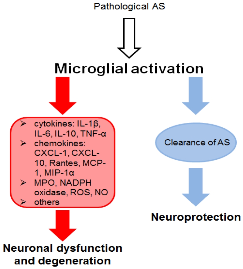
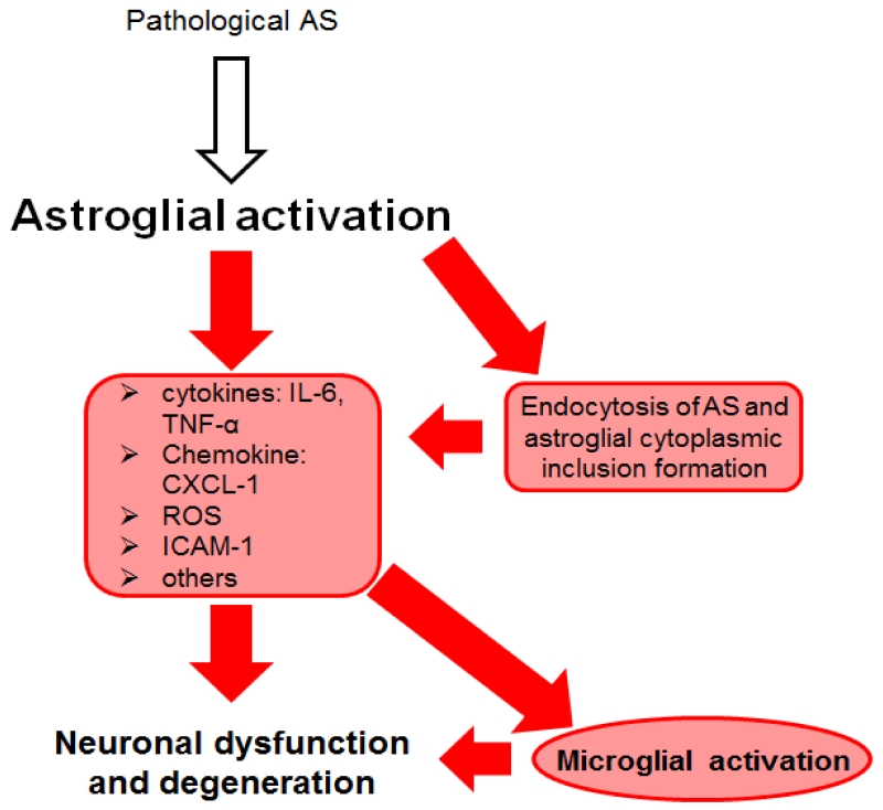
α-Synucleinopathies (ASP) comprise adult-onset, progressive neurodegenerative disorders such as Parkinson's disease (PD), dementia with Lewy bodies (DLB) and multiple system atrophy (MSA) that are characterized by α-synuclein (AS) aggregates in neurons or glia. PD and DLB feature neuronal AS-positive inclusions termed Lewy bodies (LB) whereas glial cytoplasmic inclusions (GCIs, Papp-Lantos bodies) are recognized as the defining hallmark of MSA. Furthermore, AS-positive cytoplasmic aggregates may also be seen in astroglial cells of PD/DLB and MSA brains. The glial AS-inclusions appear to trigger reduced trophic support resulting in neuronal loss. Moreover, microgliosis and astrogliosis can be found throughout the neurodegenerative brain and both are key players in the initiation and progression of ASP. In this review, we will highlight AS-dependent alterations of glial function and their impact on neuronal vulnerability thereby providing a detailed summary on the multifaceted role of glia in ASP.

Keywords: Astroglia; Dementia with Lewy bodies; Glial cytoplasmic inclusions; Lewy bodies; Microglia; Multiple system atrophy; Oligodendroglia; Parkinson's disease; α-Synuclein.


Figures

Figure 1
Figure 1. Microglial involvement in α-synucleinopathies (ASP)
Microglial cells can get activated by pathological α-synuclein (AS) (Su et al., 2009, Halliday and Stevens 2011, Fellner et al., 2013a). Different sources of these pathological AS species were proposed including release by neurons to the extracellular space or cell-to-cell propagation (Braak et al., 2007, Lee et al., 2010). Activation of microglial cells induces an oxidative stress response including the release of reactive oxygen species (ROS) and nitric oxide (NO) as well as the production of NADPH oxidase. Furthermore, pro-inflammatory cytokines, such as Interleukine-1β (IL-1β), IL-6, and tumor necrosis factor α (TNF-α), and the anti-inflammatory cytokine IL-10 as well as pro-inflammatory chemokines including (C-X-C motif) ligand 1 (CXCL-1), CXCL-10, Rantes, monocyte chemotactic protein 1 (MCP-1), macrophage inflammatory protein 1α (MIP-1α) are released by activated microglial cells (Zhang et al., 2005, Su et al., 2008, Roodveldt et al., 2010, Alvarez-Erviti et al., 2011, Rojanathammanee et al., 2011, Fellner et al., 2013a). An involvement of Toll-like receptor 4 (TLR4), TLR2 and myeloperoxidase (MPO, key enzyme related to oxidative stress during inflammation) in inflammation and oxidative stress has been suggested (Stefanova et al., 2012a, Fellner et al., 2013a, Kim et al., 2013). Inflammation and oxidative stress mediated through microglial cells can further lead to neuronal dysfunction and cell death (Zhang et al., 2005, Reynolds et al., 2008). Thereby, dying neurons might release accumulated AS that stays in the extracellular space and again leads to the activation of microglial cells. This feedback loop might increase microglial activation leading to microgliosis. However, microglial cells are also able to phagocytose different forms of extracellular AS via TLR4 (Stefanova et al., 2011, Fellner et al., 2013a). This clearance mechanism might be even beneficial for neuronal survival. The different features displayed by microglial cells make it hard to categorize the role of microglial cells in ASP. Yet, the detrimental and beneficial functions of microglial cells suggest an involvement of microglial activation in the initiation and progression of ASP (Halliday and Stevens 2011). However, further studies have to be conducted to understand the complete participation of microglial activation in ASP.
Figure 2
Figure 2. Astroglial involvement in α-synucleinopathies (ASP)
Astroglial cells are activated by different forms of α-synuclein (AS). Different sources of these pathological AS species were proposed including release by neurons or cell-to-cell propagation (Braak et al., 2007, Lee et al., 2010). Thereby AS-induced release of intercellular adhesion molecule 1 (ICAM-1), reactive oxygen species (ROS) and pro-inflammatory cytokines [e.g. Interleukine-6 (IL-6) and tumor necrosis factor α (TNF-α)] was measured (Klegeris et al., 2006, Fellner et al., 2013a). An involvement of Toll-like receptor 4 (TLR4), myeloperoxidase (MPO, key enzyme related to oxidative stress during inflammation) and hydrogen sulphide in inflammation and oxidative stress has been suggested (Choi et al., 2005, Lee et al., 2009, Fellner et al., 2013a). Furthermore, various studies found that astroglial cells can internalize extracellular or neuron-derived AS via endocytosis (Lee et al., 2010, Braidy et al., 2013, Fellner et al., 2013a). As a consequence, AS-dependent inflammation and oxidative stress and the uptake and accumulation of AS might induce microglial activation as well as neuronal dysfunction and neurodegeneration (Lee et al., 2009, Gu et al., 2010). Astroglial cells are highly involved in inflammation and neuronal cell death. Some neuroprotective features were described for astroglial cells, yet not in an AS-dependent context (Saavedra et al., 2006, Sandhu et al., 2009). Further studies have to be completed to elucidate the role of AS-endocytosis and to understand the complete picture of astroglial involvement in ASP.

References

    1. Abeliovich A, Schmitz Y, Farinas I, Choi-Lundberg D, Ho WH, Castillo PE, Shinsky N, Verdugo JM, Armanini M, Ryan A, Hynes M, Phillips H, Sulzer D, Rosenthal A. Mice lacking alpha-synuclein display functional deficits in the nigrostriatal dopamine system. Neuron. 2000;25(1):239–252. - PubMed
    1. Aerts MB, Esselink RA, Abdo WF, Bloem BR, Verbeek MM. CSF alpha-synuclein does not differentiate between parkinsonian disorders. Neurobiol Aging. 2012;33(2):430, e431–433. - PubMed
    1. Ahmed Z, Asi YT, Sailer A, Lees AJ, Houlden H, Revesz T, Holton JL. The neuropathology, pathophysiology and genetics of multiple system atrophy. Neuropathol Appl Neurobiol. 2012;38(1):4–24. - PubMed
    1. Al-Chalabi A, Durr A, Wood NW, Parkinson MH, Camuzat A, Hulot JS, Morrison KE, Renton A, Sussmuth SD, Landwehrmeyer BG, Ludolph A, Agid Y, Brice A, Leigh PN, Bensimon G, Group, N. G. S. Genetic variants of the alpha-synuclein gene SNCA are associated with multiple system atrophy. PLoS One. 2009;4(9):e7114. - PMC - PubMed
    1. Alvarez-Erviti L, Couch Y, Richardson J, Cooper JM, Wood MJ. Alpha-synuclein release by neurons activates the inflammatory response in a microglial cell line. Neurosci Res. 2011;69(4):337–342. - PubMed

Publication types

Substances