. 2016 Feb;26(2):783-796.
doi: 10.1093/cercor/bhv041. Epub 2015 Mar 12.

Attention Stabilizes Representations in the Human Hippocampus

Affiliations

Attention Stabilizes Representations in the Human Hippocampus

Mariam Aly et al. Cereb Cortex. 2016 Feb.

Abstract

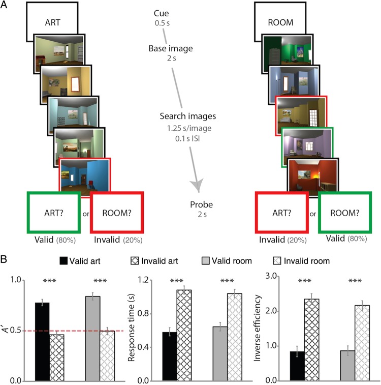
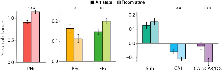
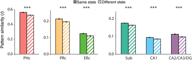
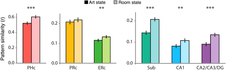
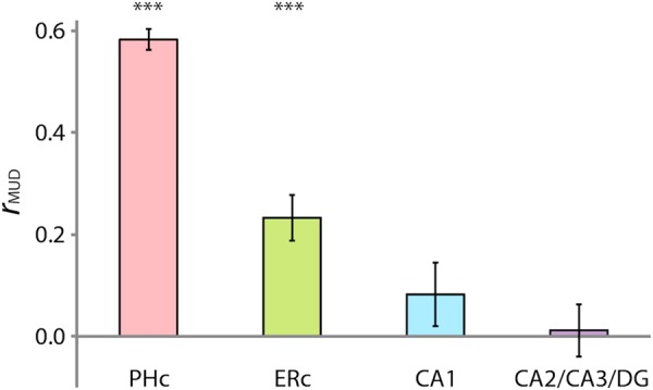
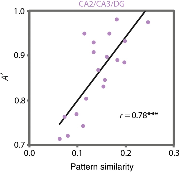
Attention and memory are intricately linked, but how attention modulates brain areas that subserve memory, such as the hippocampus, is unknown. We hypothesized that attention may stabilize patterns of activity in human hippocampus, resulting in distinct but reliable activity patterns for different attentional states. To test this prediction, we utilized high-resolution functional magnetic resonance imaging and a novel "art gallery" task. On each trial, participants viewed a room containing a painting, and searched a stream of rooms for a painting from the same artist (art state) or a room with the same layout (room state). Bottom-up stimulation was the same in both tasks, enabling the isolation of neural effects related to top-down attention. Multivariate analyses revealed greater pattern similarity in all hippocampal subfields for trials from the same, compared with different, attentional state. This stability was greater for the room than art state, was unrelated to univariate activity, and, in CA2/CA3/DG, was correlated with behavior. Attention therefore induces representational stability in the human hippocampus, resulting in distinct activity patterns for different attentional states. Modulation of hippocampal representational stability highlights the far-reaching influence of attention outside of sensory systems.

Keywords: attentional modulation; high-resolution fMRI; hippocampal subfields; medial temporal lobe; task representations.


Figures

Figure 1.
Figure 1.
Behavioral task. Two sample trials are depicted (see text for details about task instructions). For visualization, cued matches are outlined in green and uncued matches in red. (A) Example of an art-state trial, with cued match absent and uncued match present, and a room-state trial, with cued match present and uncued match present. (B) Sensitivity, RT, and inverse efficiency in making present/absent judgment as a function of attentional state and probe type. Error bars depict ±1 SEM of the within-subject valid versus invalid difference. Dashed line indicates chance performance. ***P < 0.001.
Figure 2.
Figure 2.
MTL ROIs. Example segmentation from one participant is depicted for an anterior and a posterior slice. ROIs consisted of 3 hippocampal subfields (subiculum [Sub], CA1, and CA2/CA3/DG) and 3 MTL cortical regions (PRc, ERc, and PHc). For segmentation guide, see Supplementary Methods.
Figure 3.
Figure 3.
Attentional modulation of univariate activity. BOLD activity evoked in the art and room states was extracted from all voxels in each ROI and averaged. Baseline corresponds to unmodeled periods of passive viewing of a blank screen. In MTL cortex, PHc and ERc were more active in the room state and PRc was more active in the art state. In the hippocampus, CA1 and CA2/CA3/DG were also more active in the art state (or deactivated in the room state). Error bars depict ±1 SEM of the within- subject art versus room state difference. *P < 0.05, **P < 0.01, ***P < 0.001.
Figure 4.
Figure 4.
State-dependent pattern similarity. BOLD activity evoked in the art and room states was extracted from all voxels in each ROI and correlated across trials of the same versus different states. In MTL cortex, all regions showed greater pattern similarity for same versus different states. In the hippocampus, all subfields showed greater pattern similarity for same versus different states. Results are shown as Pearson correlations, but statistical tests were performed only after applying the Fisher transformation. Error bars depict ±1 SEM of the within-subject same versus different state difference. ***P < 0.001.
Figure 5.
Figure 5.
Comparison of pattern similarity between states. BOLD activity was extracted from all voxels in each ROI and separately correlated across trials of the art and room states, respectively. In MTL cortex, PHc and ERc showed greater pattern similarity for room versus art states, and PRc showed no difference. In the hippocampus, all subfields showed greater pattern similarity for room versus art states. Results are shown as Pearson correlations, but statistical tests were performed only after applying the Fisher transformation. Error bars depict ±1 SEM of the within-subject art- versus room-state difference. **P < 0.01, ***P < 0.001.
Figure 6.
Figure 6.
Multivariate-univariate dependence (MUD) analysis. The contribution of each voxel to pattern similarity was estimated by normalizing BOLD activity over voxels within an ROI for each trial and computing pairwise products across trials. Average products from room trials were then correlated with average activity in room trials over voxels to estimate MUD. In MTL cortex, PHc and ERc showed a positive relationship between activity and pattern similarity. In the hippocampus, CA1 and CA2/CA3/dentate gyrus (DG) showed no relationship. Error bars depict ±1 SEM across participants. Results are shown as Pearson correlations, but statistical tests were performed only after applying the Fisher transformation. ***P < 0.001.
Figure 7.
Figure 7.
Brain–behavior relationships. Individual differences in room-state pattern similarity in CA2/CA3/DG were strongly correlated with individual differences in behavioral performance (A′) on the room task. This effect was specific to this region, to the room task, and to the pattern similarity measure. ***P < 0.001.

References

    1. Adcock RA, Thangavel A, Whitfield-Gabrieli S, Knutson B, Gabrieli JDE. 2006. Reward-motivated learning: mesolimbic activation precedes memory formation. Neuron. 50:507–517. - PubMed
    1. Aly M, Ranganath C, Yonelinas AP. 2013. Detecting changes in scenes: the hippocampus is critical for strength-based perception. Neuron. 78:1127–1137. - PMC - PubMed
    1. Bonnici HM, Kumaran D, Chadwick MJ, Weiskopf N, Hassabis D, Maguire EA. 2012. Decoding representations of scenes in the medial temporal lobes. Hippocampus. 22:1143–1153. - PMC - PubMed
    1. Brown MW, Aggleton JP. 2001. Recognition memory: what are the roles of the perirhinal cortex and hippocampus? Nat Rev Neurosci. 2:51–61. - PubMed
    1. Brown TI, Ross RS, Keller JB, Hasselmo ME, Stern CE. 2010. Which way as I going? Contextual retrieval supports the disambiguation of well learned overlapping navigational routes. J Neurosci. 30:7414–7422. - PMC - PubMed

Publication types