. 2015 Jan-Mar;10(1):47.
doi: 10.4103/1793-5482.151513.

Microsurgical management of aneurysms of the superior cerebellar artery - lessons learnt: An experience of 14 consecutive cases and review of the literature

Affiliations

Microsurgical management of aneurysms of the superior cerebellar artery - lessons learnt: An experience of 14 consecutive cases and review of the literature

Prakash Nair et al. Asian J Neurosurg. 2015 Jan-Mar.

Abstract

Objective: This is a retrospective study from January 2002 to December 2012 analyzing the results of microsurgical clipping for aneurysms arising from the superior cerebellar artery (SCA).

Materials and methods: All patients with SCA were evaluated with computerized tomography angiography and/or digital subtraction angiography (DSA) prior to surgery. All patients in our series underwent microsurgical clipping and postoperative DSA to assess the extent of aneurysm occlusion. The Glasgow outcome scale (GOS) and the modified Rankin's scale (mRS) were used to grade their postoperative neurological status at discharge and 6 months, respectively.

Results: Fourteen patients had SCA aneurysms (ruptured-9, unruptured-5). There were 10 females and 4 males with the mean age of 47.2 years (median - 46 years, range = 24-66 years). Subarachnoid hemorrhage (SAH) was seen in 11 patients. The mean duration of symptoms was 2.5 days (range = 1-7 days). The WFNS score at presentation was as follows: Grade 1 in 10 cases, II in 2 cases, III in 1 case and IV in 1 case. In the 9 cases with ruptured SCA aneurysm, average size of the ruptured aneurysms was 7.3 mm (range = 2.5-27 mm, median = 4.9 mm). The subtemporal approach was used in the first 7 cases. The extradural temporopolar (EDTP) approach was used in the last 5 cases. Complications include vasospasm (n = 6), third nerve palsy (n = 5) and hydrocephalus (n = 3). Two patients died following surgery. At mean follow-up 33.8 months (median - 25 months, range = 19-96 months), no patient had a rebleed. At discharge 9 (64%), had a GOS of 4 or 5 and 3 (21%) had a GOS of 3. At 6 months follow-up, 10/14 (71%) patients had mRS of 0-2, and 2 (14%) had mRS of 5.

Conclusions: Aneurysms of the SCA are uncommon and tend to rupture even when the aneurysm size is small (<7 mm). They commonly present with SAH. The EDTP approach avoids complication caused by temporal lobe retraction and injury to the vein of Labbe.

Keywords: Accessory superior cerebellar artery; aneurysm; extradural temporopolar approach; subarachnoid hemorrhage; superior cerebellar artery.


Conflict of interest statement

Conflict of Interest: None declared.

Figures

Figure 1
Figure 1
(a-c) A 34-year-old female patient presented with severe acute headache and vomiting of 2 days duration. DSA done shows a 3.8 mm aneurysm arising from the right SCA, just distal to its origin from the basilar artery, pointing upward and posteriorly. (d) She underwent aneurysm clipping by subtemporal approach. Postoperative DSA showed complete aneurysm occlusion and preserved flow in the distal SCA
Figure 2
Figure 2
(a) CT showed SAH with a hematoma in the left temporal lobe in a 45-year-old male seen in the emergency with sudden onset headache and loss of consciousness. (b) DSA showing a left MCA and ICA aneurysm, and (c) shows a superiorly directed SCA aneurysm. (d) Operative pictures seen through a subtemporal approach with the aneurysm arising from the SCA, the oculomotor nerve and PCA are seen in close relation. (e) The final clip placement across the aneurysm, and (f) DSA showing complete occlusion of the aneurysm
Figure 3
Figure 3
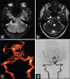
(a and b) MRI of a patient with trigeminal neuralgia, a T2 hypointense lesion is seen in the right cerebellopontine angle, the susceptility weighted imaging shows evidence of blood (c) Computerized tomography angiogram shows a partially thrombosed distal right SCA aneurysm with a left sided PCA aneurysm. (d) Digital subtraction angiography shows only partial filling right SCA aneurysm, the left PCA aneurysm appears to be fusiform involving the main vessel. The SCA aneurysm was trapped using a retromastoid approach; the PCA aneurysm was coiled later
Figure 4
Figure 4
(a) A 38-year-old women presented with SAH in the interpeduncular cistern. Initial DSA showed no aneurysm. (b-d) Computerized tomography angiogram and DSA done 6 weeks later showed a fusiform aneurysm arising from an accessory superior cerebellar artery (aSCA), which took origin from the basilar artery distal to the main SCA (e and f) Intraoperative pictures of the surgical field through a temporopolar approach, showing a fusiform aneurysm arising from the aSCA, which was then trapped (BA: Basilar artery, SCA: Superior cerebellar artery, aSCA: Accessory superior cerebellar artery, III - oculomotor nerve)
Figure 5
Figure 5
(a) CTA and (b and c) DSA of a 66-year-old female with Grade 3 SAH showing a 4.9 mm aneurysm arising from the left SCA, directed upward and posteriorly. The right PCA and SCA are seen arising from a single common trunk. The aneurysm was clipped using a temporopolar approach. (d) The aneurysm arising from the SCA, distal to its origin. (e) And the final clip placement across the aneurysm neck. (f) Postoperative angiogram shows complete occlusion of the aneurysm and flow across the patent SCA)
Figure 6
Figure 6
(a) A 28-year-old man with SAH was found to have an upward directed aneurysm arising in the distal SCA (open arrow), which was approached by the temporopolar approach. (b) Postoperative angiogram after clipping shows complete occlusion of the aneurysm neck and flow through the parent vessel (closed arrow)

Similar articles

Cited by

References

    1. Locksley HB. Natural history of subarachnoid hemorrhage, intracranial aneurysms and arteriovenous malformations. J Neurosurg. 1966;25:321–68. - PubMed
    1. Peluso JP, van Rooij WJ, Sluzewski M, Beute GN. Superior cerebellar artery aneurysms: Incidence, clinical presentation and midterm outcome of endovascular treatment. Neuroradiology. 2007;49:747–51. - PMC - PubMed
    1. Chaloupka JC, Putman CM, Awad IA. Endovascular therapeutic approach to peripheral aneurysms of the superior cerebellar artery. AJNR Am J Neuroradiol. 1996;17:1338–42. - PMC - PubMed
    1. Gács G, Viñuela F, Fox AJ, Drake CG. Peripheral aneurysms of the cerebellar arteries. Review of 16 cases. J Neurosurg. 1983;58:63–8. - PubMed
    1. Jin SC, Park ES, Kwon do H, Ahn JS, Kwun BD, Kim CJ, et al. Endovascular and microsurgical treatment of superior cerebellar artery aneurysms. J Cerebrovasc Endovasc Neurosurg. 2012;14:29–36. - PMC - PubMed