Case Reports
. 2015 Jan-Mar;10(1):55.
doi: 10.4103/1793-5482.151522.

Contralateral transvenous approach and embolization with 360° guglielmi detachable coils for the treatment of cavernous sinus dural fistula

Affiliations
Case Reports

Contralateral transvenous approach and embolization with 360° guglielmi detachable coils for the treatment of cavernous sinus dural fistula

Marco Zenteno et al. Asian J Neurosurg. 2015 Jan-Mar.

Abstract

carotid-cavernous fistulas are spontaneours acquired connections between the carotid artery and the cavernous cavernous sinus, being classified as direct or indirect; being usually diagnosed in postmenopausal women, but are also associated with other pathoogies such as pregnancy, sinusitis and cavernous sinus thrombosis. They are clinically characterized by ophthalmological symptoms and pulsatile tinnitus. A 51-year-old woman who started her current condition about 4 years ago with pulsatile tinnitus, to which were added progressively: Pain, conjunctival erythema, right eye proptosis and the occasional headache of moderate intensity. Caotid-cavernous fistula wes diagnosed, for the technical difficulty inherent in the case was made a contralateral transvenous approach and embolization with 360° GDG coils, with successful evolution of the patient. The endovascular management of these lesions is currently possible with excellent results.

Keywords: Cavernous sinus; dural fistula; neurointerventional therapy.


Conflict of interest statement

Conflict of Interest: None declared.

Figures

Figure 1
Figure 1
It is shown the right proptosis and bipalpebral edema (a). It also can be observed the congestion of conjunctival vessels in a “caput medusae” and “corkscrew” fashion (b and c)
Figure 2
Figure 2
T2-weighted magnetic resonance imaging (MRI) shows the right eye propotosis (a), the ingurgitation of orbital veins and edema of the extraocular muscles (b, arrows). Note the dilatation and the absence of signal due to high-flow in the left cavernous sinus (c and d, arrowheads) and in the circular sinus toward the right cavernous sinus (c and d, hollow arrows)
Figure 3
Figure 3
Digital subtraction (a: Lateral view, b: Left oblique view, and c: Later view and more lateralized than b) and selective three-dimensional (d: AP view) angiography of the left internal carotid artery. It is seen the pass to the left cavernous sinus through meningeal branches of the meningohipofisarial and inferolateral trunks (b and d, thin arrows). The contrast material passes through intercavernous sinus (b and d, arrows) into the right cavernous sinus (a, b and d, arrowheads). Retrograde flow is observed from the inferiro petrosal sinus (a, b, c and d, thick arrow) and the ophthalmic vein (c, star)
Figure 4
Figure 4
Digital subtraction (a: Lateral view, b: Lateral view later stage than a and c: AP view) and selective three-dimensional (d: Anterolateral view) angiography of the left external carotid artery. It is observed the passage of contrast material through direct branches of the maxillary artery and in turn the middle meningeal artery toward the left cavernous sinus (a, c, d, thin arrows). The contrast material passes to the contralateral cavernous sinus (a-d, arrowhead) through intercavernous sinus (c and d, arrows). Retrograde flow can be seen through the inferior petrosal sinus (a-d, thick arrow) and the ophthalmic vein (b, star)
Figure 5
Figure 5
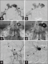
Fistula management by contrast injection through arterial approach, where there are visualized the tributary vessels (a, thick arrow) and venous embolization. The distal end of the guide catheter was fixed at the level of the internal jugular vein (a, b and c: Arrowhead), and through this was passed a microcatheter that coursed the inferior petrosal sinus, the right cavernous and intercavernous sinuses (a-d and f, thin arrows). The distal end of the microcatheter was placed in the left cavernous sinus (b, arrow) and was started the coils placing and releasing (c-f, Star). By the end of angiographic embolization, meningeal vessels were observed, with no communication with the cavernous sinus (e, thick arrow)
Figure 6
Figure 6
Photograph of AO 1 week after treatment. The proptosis, bipalpebral edema, and congestion of conjunctival vessels are no longer observed (a and b). Eye movements are conserved (c-e)
Figure 7
Figure 7
Magnetic resonance imaging 15 days after endovascular treatment. At orbital level is no longer observed extraocular muscle edema or bulking ophthalmic veins (a). The tetralogy of fallot angio magnetic resonance imaging shows absence of the fistula (b). Can be appreciated hypointensity originated by coils within the left cavernous and intercoronary sinuses (c and d, arrows), besides the signal absence of blood flow within the internal carotid artery (c and d, arrows)

Similar articles

References

    1. Debrun GM, Viñuela F, Fox AJ, Davis KR, Ahn HS. Indications for treatment and classification of 132 carotid-cavernous fistulas. Neurosurgery. 1988;22:285–9. - PubMed
    1. Jacobson BE, Nesbit GM, Ahuja A, Barnwell SL. Traumatic indirect carotid-cavernous fistula: Report of two cases. Neurosurgery. 1996;39:1235–7. - PubMed
    1. Meyers PM, Halbach VV, Dowd CF, Lempert TE, Malek AM, Phatouros CC, et al. Dural carotid cavernous fistula: Definitive endovascular management and long-term follow-up. Am J Ophthalmol. 2002;134:85–92. - PubMed
    1. Yamashita K, Taki W, Nishi S, Sadato A, Nakahara I, Kikuchi H, et al. Transvenous embolization of dural caroticocavernous fistulae: Technical considerations. Neuroradiology. 1993;35:475–9. - PubMed
    1. Higashida RT, Halbach VV, Tsai FY, Norman D, Pribram HF, Mehringer CM, et al. Interventional neurovascular treatment of traumatic carotid and vertebral artery lesions: Results in 234 cases. AJR Am J Roentgenol. 1989;153:577–82. - PubMed

Publication types