. 2015 Mar 13;10(3):e0120647.
doi: 10.1371/journal.pone.0120647. eCollection 2015.

CCAAT/enhancer binding protein β is dispensable for development of lung adenocarcinoma

Affiliations

CCAAT/enhancer binding protein β is dispensable for development of lung adenocarcinoma

Yi Cai et al. PLoS One. .

Abstract

Lung cancer is the leading cause of cancer death worldwide. Although disruption of normal proliferation and differentiation is a vital component of tumorigenesis, the mechanisms of this process in lung cancer are still unclear. A transcription factor, C/EBPβ is a critical regulator of proliferation and/or differentiation in multiple tissues. In lung, C/EBPβ is expressed in alveolar pneumocytes and bronchial epithelial cells; however, its roles on normal lung homeostasis and lung cancer development have not been well described. Here we investigated whether C/EBPβ is required for normal lung development and whether its aberrant expression and/or activity contribute to lung tumorigenesis. We showed that C/EBPβ was expressed in both human normal pneumocytes and lung adenocarcinoma cell lines. We found that overall lung architecture was maintained in Cebpb knockout mice. Neither overexpression of nuclear C/EBPβ nor suppression of CEBPB expression had significant effects on cell proliferation. C/EBPβ expression and activity remained unchanged upon EGF stimulation. Furthermore, deletion of Cebpb had no impact on lung tumor burden in a lung specific, conditional mutant EGFR lung cancer mouse model. Analyses of data from The Cancer Genome Atlas (TCGA) revealed that expression, promoter methylation, or copy number of CEBPB was not significantly altered in human lung adenocarcinoma. Taken together, our data suggest that C/EBPβ is dispensable for development of lung adenocarcinoma.


Conflict of interest statement

Competing Interests: D. B. C. has previously received consulting fees and honoraria from Pfizer and S. S. K. has previously received consulting fee from Bristol-Myers Squibb. The authors declare that this does not alter their adherence to all PLOS ONE policies on sharing data and materials. No other authors have competing interests.

Figures

Fig 1
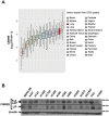
Fig 1. C/EBPβ is expressed in human lung cells.
(A) Expression of CEBPB in various human tissues in the dataset from GTEx project. RPKM values were log2 transformed and presented as a box plot for each tissue type. (B) Protein extracts were isolated from a panel of human lung adenocarcinoma cell lines as well as an immortalized human bronchoepithelial cell line, BEAS-2B and subjected to Western blotting.
Fig 2
Fig 2. Deletion of Cebpb has no effect on lung structure, but leads to aggregation of lymphoid cells in the lung.
(A) Immunoblots of lung extracts from Cebpb +/+ or Cebpb -/- mice. Note that the predominant isoform expressed in mouse lungs is LAP and C/EBPβ expression in the lung is effectively lost in Cebpb -/- mice. (B) Representative images of hematoxylin and eosin staining of lungs isolated from Cebpb +/+ (I and II) and Cebpb -/- (III and IV) mice. We analyzed 10 Cebpb +/+ and 8 Cebpb -/- mice. Scale bars = 500 μm (I and III) and 100 μm (II and IV). Arrows indicate scattered lymphocyte aggregates in the lung parenchyma of a Cebpb -/- mouse. No gross or histologic architectural abnormalities were observed in the lungs of Cebpb -/- mice.
Fig 3
Fig 3. C/EBPβ fails to contribute to cell proliferation in vitro.
(A) Fluorescent images of NCI-H358 cells stably expressing C/EBPβ-ER. Cells were treated with 1 μM β-estradiol or 0.1% ethanol (vehicle) for 24 hours and stained with anti ER antibody and visualized by Alexa Fluor 488. Scale Bar = 10 μm. (B) Luciferase assay to determine C/EBPβ transcriptional activity in NCI-H358 cells stably expressing C/EBPβ-ER and control cells. Cells were transfected with C/ebpb0.3-luc reporter and pCMV-Renilla control plasmid were assayed after 24 hours of treatment with 1 μM β-estradiol or vehicle control. Data represents mean ± standard deviation from three independent experiments. ** denotes p ≤ 0.01. (C) Cell viability determined by MTS assay. NCI-H358 cells expressing C/EBPβ-ER cells or control cells were plated in 96 well plates at 2,000 cells/well and assayed after 72 hours of incubation. Data represents mean ± standard deviation from eight independent experiments. * denotes p ≤ 0.05. (D) Immunoblots of C/EBPβ in NCI-H358 cells transduced with shRNA against CEBPB. Three clones each for control and shRNA against CEBPB are shown. (E) Cell viability determined by MTS assay. NCI-H358 cells transduced with shRNA against CEBPB or control shRNA described in (D) were plated in 96 well plates at 2,000 cells/well and assayed after 72 hours of incubation. Data represents mean ± standard deviation from three independent experiments. N.S. denotes not significant.
Fig 4
Fig 4. EGF does not increase LIP protein.
(A) Immunoblots of C/EBPβ in NCI-H1975 (EGFR L858R+T790M) and A549 (KRAS G12S) lung adenocarcinoma cells treated with EGF in the presence or absence of EGFR kinase inhibitors for 24 hours. Changes in LIP expression are adjusted by β-actin expression and expressed as fold increase relative to that of PBS-treated cells. Note that the full and LAP forms were detected in the gel exposed for 15 seconds, whereas LIP forms were detected after 1-hourr exposure of the gel. (B) Luciferase assay to determine C/EBPβ transcriptional activity. NCI-H1975 and A549 cells transfected with C/ebpb0.3-luc reporter and pCMV-Renilla control plasmid were assayed after 24 hours of treatment with EGF or PBS control. Data represents mean ± standard deviation from four independent experiments. N.S. denotes not significant.
Fig 5
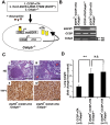
Fig 5. Deletion of Cebpb does not affect tumor burden in vivo.
(A) Scheme of the strategy generating lung specific inducible EGFR-TL transgenic mice with Cebpb knockout background. (B) Genotype of EGFR TL /CCSP-rtTA/Cebpb +/+, CCSP-rtTA/Cebpb +/+ and EGFR TL /CCSP-rtTA/Cebpb -/- mice. (C) Representative images of H&E staining and immunohistochemistry of lungs isolated from EGFR TL /CCSPrtTA/Cebpb +/+(I and II) or EGFR TL /CCSPrtTA/Cebpb -/-(III and IV). Lung sections were stained with H&E (I and III) and anti-TTF1 (II and IV) antibodies. “A” indicates an area with bronchioalveolar growth pattern and “P” indicates an area with bronchial papillary growth pattern. (D) Lung weight isolated from mice treated with doxycycline for 8–12 weeks. We analyzed 5 CCSP-rtTA/Cebpb +/+, 5 EGFR TL /CCSP-rtTA/Cebpb +/+, and 11 EGFR TL /CCSP-rtTA/Cebpb -/- mice. Data represent mean ± standard deviation. ** denotes p ≤ 0.01. N.S. denotes not significant.
Fig 6
Fig 6. C/EBPβ is not altered in human lung adenocarcinoma.
(A) mRNA expression of CEBPA, CEBPB, and CDKN2A in TCGA lung adenocarcinoma dataset. RSEM values obtained from TCGA data were log2 transformed and depicted as individual dots representing a sample and box plot. (B) Methylation status in CEBPA and CEBPB promoter regions in TCGA lung adenocarcinoma dataset. Scatter plot depicts mRNA expression in Y-axis and methylation level of the promoter region in X-axis of CEBPA and CEBPB genes. (C) Copy number estimates of CEBPA and CEBPB gene loci in TCGA lung adenocarcinoma dataset. Scatter plot depicts mRNA expression in Y-axis and copy number estimates in log2 scale in X-axis of CEBPA and CEBPB genes.

Similar articles

Cited by

References

    1. Siegel R, Ma J, Zou Z, Jemal A. Cancer statistics, 2014. CA: a cancer journal for clinicians. 2014;64(1):9–29. - PubMed
    1. Cersosimo RJ. Lung cancer: a review. American journal of health-system pharmacy: AJHP: official journal of the American Society of Health-System Pharmacists. 2002;59(7):611–42. - PubMed
    1. Dela Cruz CS, Tanoue LT, Matthay RA. Lung cancer: epidemiology, etiology, and prevention. Clinics in chest medicine. 2011;32(4):605–44. 10.1016/j.ccm.2011.09.001 - DOI - PMC - PubMed
    1. Herceg Z, Vaissiere T. Epigenetic mechanisms and cancer: an interface between the environment and the genome. Epigenetics: official journal of the DNA Methylation Society. 2011;6(7):804–19. 10.4161/epi.6.7.16262 - DOI - PubMed
    1. Liloglou T, Bediaga NG, Brown BR, Field JK, Davies MP. Epigenetic biomarkers in lung cancer. Cancer letters. 2014;342(2):200–12. 10.1016/j.canlet.2012.04.018 - DOI - PubMed

Publication types