Review
. 2015 Mar 11;16(3):5635-65.
doi: 10.3390/ijms16035635.

Molecular mechanisms underlying β-adrenergic receptor-mediated cross-talk between sympathetic neurons and immune cells

Affiliations
Review

Molecular mechanisms underlying β-adrenergic receptor-mediated cross-talk between sympathetic neurons and immune cells

Dianne Lorton et al. Int J Mol Sci. .

Abstract

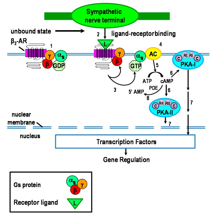
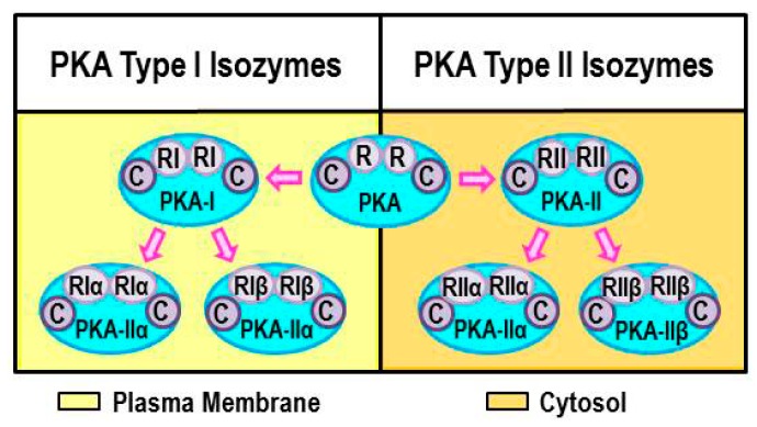
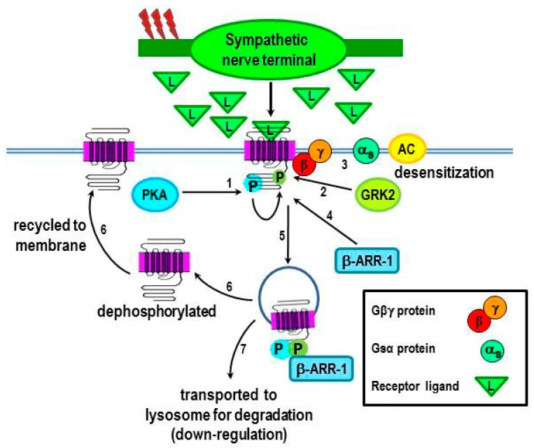
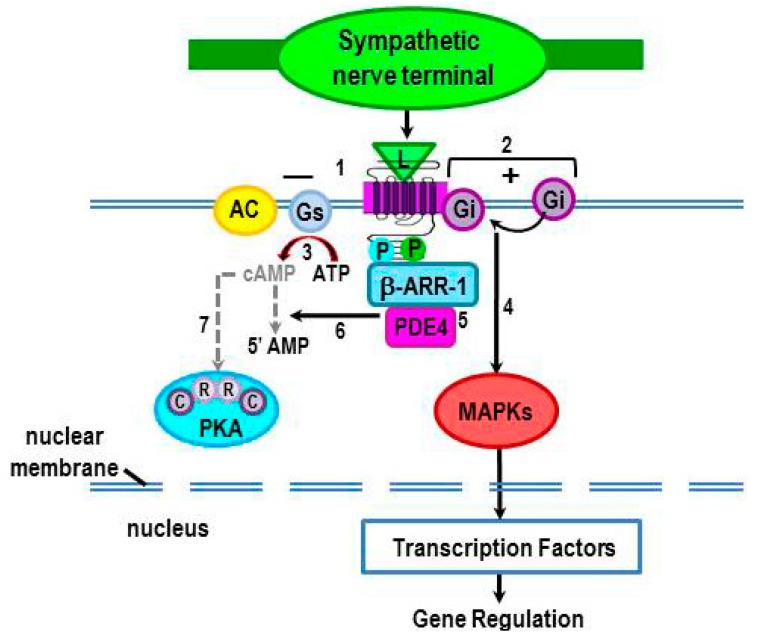
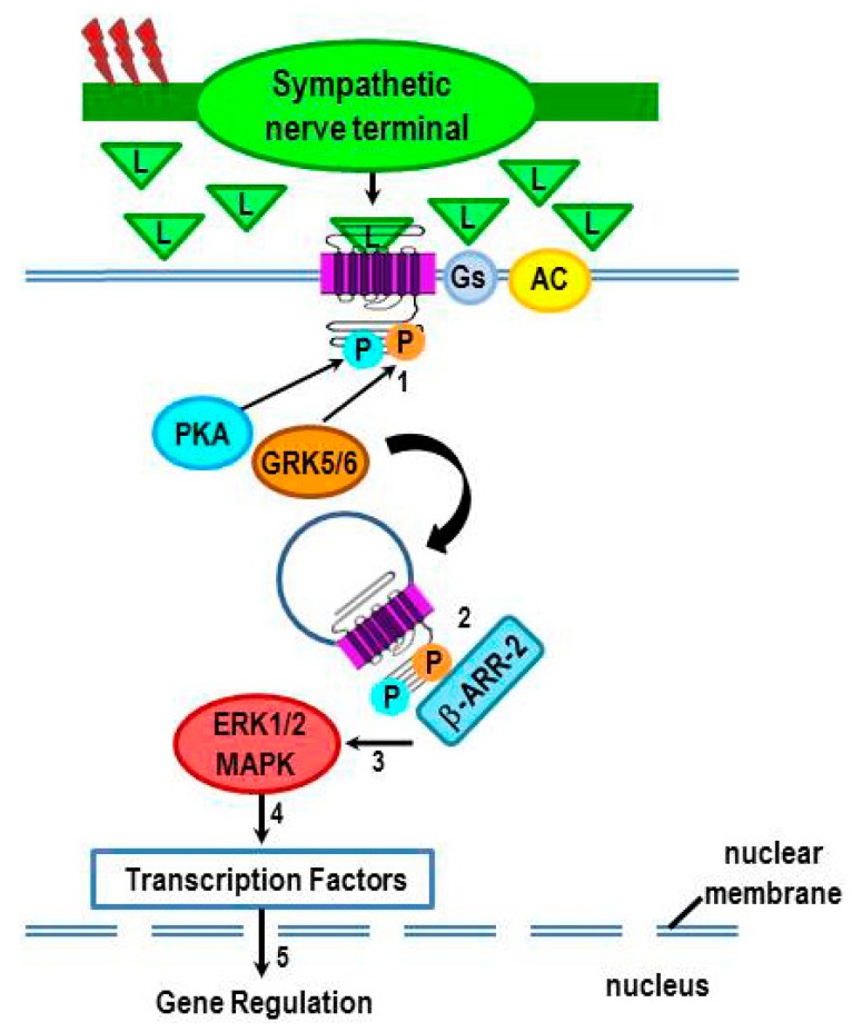
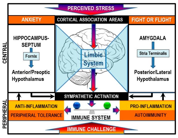
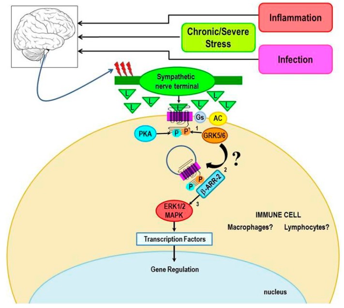
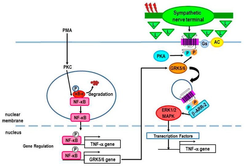
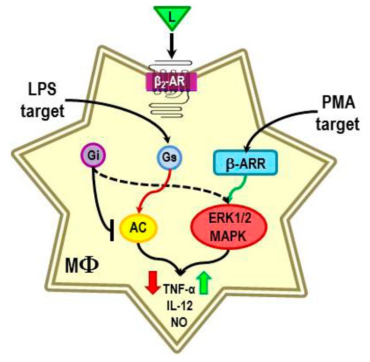
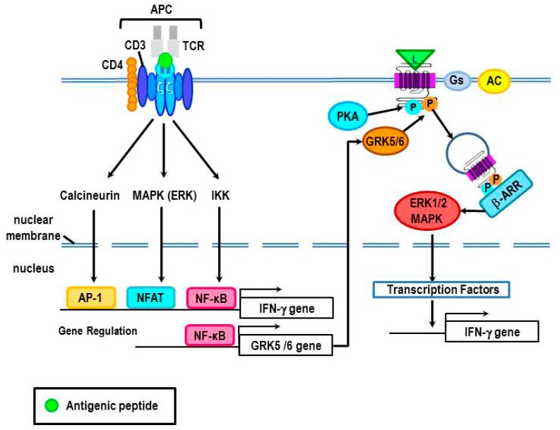
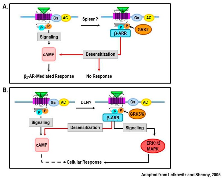
Cross-talk between the sympathetic nervous system (SNS) and immune system is vital for health and well-being. Infection, tissue injury and inflammation raise firing rates of sympathetic nerves, increasing their release of norepinephrine (NE) in lymphoid organs and tissues. NE stimulation of β2-adrenergic receptors (ARs) in immune cells activates the cAMP-protein kinase A (PKA) intracellular signaling pathway, a pathway that interfaces with other signaling pathways that regulate proliferation, differentiation, maturation and effector functions in immune cells. Immune-SNS cross-talk is required to maintain homeostasis under normal conditions, to develop an immune response of appropriate magnitude after injury or immune challenge, and subsequently restore homeostasis. Typically, β2-AR-induced cAMP is immunosuppressive. However, many studies report actions of β2-AR stimulation in immune cells that are inconsistent with typical cAMP-PKA signal transduction. Research during the last decade in non-immune organs, has unveiled novel alternative signaling mechanisms induced by β2-AR activation, such as a signaling switch from cAMP-PKA to mitogen-activated protein kinase (MAPK) pathways. If alternative signaling occurs in immune cells, it may explain inconsistent findings of sympathetic regulation of immune function. Here, we review β2-AR signaling, assess the available evidence for alternative signaling in immune cells, and provide insight into the circumstances necessary for "signal switching" in immune cells.


Figures

Figure 1
Figure 1
The canonical β2-adrenergic receptor (AR) signaling pathway is illustrated here. In target tissues, activated sympathetic nerves release the neurotransmitter, norepinephrine (NE) from “boutons en passage”. NE and epinephrine from the circulation are the natural ligands (L) for the β2-AR. β2-ARs couple with stimulatory Gαs subunit to modulate the activity of adenylate cyclase (AC). Receptor activation causes dissociation of Gαs from the Gβγ subunit complex of the Gs protein, which results in AC-mediated production of cAMP from ATP. Next, cAMP activates either protein kinase A-I (PKA-I) or -II. PKA activation leads to the activation of transcription factors such as cAMP response element-binding (CREB) protein to regulate gene transcription.
Figure 2
Figure 2
Illustration of the structure and current nomenclature for protein kinase A (PKA) Isoforms. Isoforms of PKA differ in the regulatory proteins (R) they express, either regulator protein I (RI) or II (RII) for PKA-I or II isoforms. PKA-I associates with the plasma membrane, whereas PKA-II localizes to cytosol and the membranes of cell organelles. Proteins called A-kinase anchor proteins (AKAPs) that bind PKAs are responsible for the site-specific localization of PKA isoforms.
Figure 3
Figure 3
Receptor responsiveness to β2-AR ligands is regulated by protein kinases that phosphorylate the receptor. PKA that is activated after β2-AR activation by ligand (L) subsequently phosphorylates the receptor (#1), which uncouples Gαs from the Gβγ subunit, this results in an uncoupling of Gαs from the receptor (#3), thus terminating receptor signaling. Phosphorylation of the receptor by PKA also facilitates receptor phosphorylation by GRK2 (#2), which further drives receptor desensitization (#3). GRK2 recruits β-arrestin-1 to the receptor (#4), a step that leads to receptor internalization (#5). The internalized β2-AR is then transported to lysosomes for degradation (#7), receptor down-regulation or dephosphorylation and recycled to the cell membrane (#5–6). Chronic stimulation of the receptor or high ligand concentrations promote the transport of the receptor to lysosomes for degradation, resulting in receptor down-regulation. Red lightning bolts indicate increased sympathetic nerve firing.
Figure 4
Figure 4
Phosphorylation of β2-ARs by PKA induces a conformational change that impairs Gs binding (#1), but enhances Gi binding (#2) to the receptor. This transient shift in receptor coupling prevents activation of AC and thus, inhibits signaling via cAMP (#3). Coupling of Gi to the receptor results in activation of mitogen-activated protein kinase (MAPKs) (#4). MAPKs subsequently phosphorylate transcription factors that regulate gene expression, including the activation of critical genes that drive innate and adaptive immunity. Binding of β-arrestin to the PKA and G-protein coupled receptor kinase 2 (GRK2)-phosphorylated receptor recruits phosphodiesterase (PDE) 4 to the receptor (#5). PDE4 degrades cAMP (#6), resulting in a reduction of local levels of cAMP, thus terminating cAMP activation of PKA (#7).
Figure 5
Figure 5
Phosphorylation of β2-AR by GRK-2, -5, and -6 occurs in an agonist concentration-dependent manner to induce different receptor functions. High sympathetic firing activity (red lightning bolts) drives the β2-AR-Gs-independent (non-canonical) pathway by flooding the extracellular space with ligand (L, i.e. NE). Chronic receptor activation induces not only PKA-mediated phosphorylation of the receptor, but also phosphorylation by GRK5/6 (#1) instead of GRK2. Receptor phosphorylation by PKA and GRK5/6 promote receptor desensitization and internalization by recruiting β-arrestin 2 (β-ARR-2) to the receptor (#2). Once bound to the receptor, β-arrestin-2 acts as a scaffold for the sustained activation of the MAPK, ERK 1/2 (#3). Beta-arrestin activation of ERK1/2 MAPK, in turn, increases the translocation of transcription factors (#4) into the nucleus to influence gene transcription (#5).
Figure 6
Figure 6
The brain perceives immune stimuli as a stressor via both humoral and via hardwired pathways to the central nervous system (CNS). This information, along with other psychosocial stressors is integrated by cortical association areas, the hypothalamus, and limbic circuits and provides a coordinated stress responses by the two major efferent stress pathways, one of which is the sympathetic nervous system (SNS). The SNS response influences the inflammatory state. Under conditions where the antigen is not eliminated and there is a break in immune tolerance autoimmunity can be triggered.
Figure 7
Figure 7
The hypothetical model for shifting β2-AR signaling from the canonical pathway towards the non-canonical pathway in immune cells is illustrated. We propose that signal transduction switching towards β-arrestin-mediated signaling occurs under conditions of unchecked immune cell activation or chronic or severe stress, both of which increase the firing rates of sympathetic nerves (red lightning bolts) and elevates local NE concentrations. These conditions favor the phosphorylation of the receptor by GRK 5/6 rather than GRK 2, thus promoting β-arrestin-2-mediated signaling via the MAPK, ERK1/2.
Figure 8
Figure 8
The presence of a β2-AR ligand during lipopolysaccharide (LPS) or phorbol 12-myristate 13-acetate (PMA)-induced macrophage activation causes reduced or increased production of TNF-α, IL-12 and nitric oxide respectively. The β2-AR-induced immunosuppression in LPS-treated macrophages is consistent with the activation of adenylyl cyclase (AC), and subsequently PKA. LPS promotes adenylyl cyclase activity, suppresses translocation of GRK2 to the membrane, and reduces the expression of GRKs 5 and 6. These LPS-induced effects are expected to increase β2-AR signaling via the cAMP-PKA canonical pathway. In contrast, the β2-AR-induced immune enhancement in PMA-treated macrophages is not consistent with receptor activation of the cAMP-PKA pathway. Instead, we propose that the responsible mechanism is a shift in receptor signaling from the canonical to the non-canonical pathway.
Figure 9
Figure 9
A possible mechanism for β2-AR-induced increases in TNF-α production in PMA-treated macrophages is by increasing gene transcription of GRK 5 or 6. PMA induces an increase in protein kinase C (PKC), which activates NF-κB by phosphorylating IκBα. Phosphorylation of IκBα releases NF-κB for nuclear translocation, and subsequent DNA binding. NF-κB increases gene expression of both TNF-α and GRK5. Increased GRK5 promotes signaling of β2-ARs in a β-arrestin-dependent manner, leading to activation of ERK1/2. ERK1/2 activates transcription factors that further promote TNF-α gene transcription to increase the production of TNF-α.
Figure 10
Figure 10
A proposed mechanism for β2-AR-mediated increases in IFN-γ in Th0 cells after antigen challenge is illustrated. T cell receptor (TCR) signal transduction is induced by the recognition of the antigen that is presented to the T cell by an antigen-presenting cell (APC). Antigen presentation can induce TCR activation of the signal transduction pathways, calcineurin, MAPK (ERK), and/or IKK, each with specific downstream signal cascades that lead to nuclear translocation of activator protein 1 (AP-1), nuclear factor of activated T cells (NFAT) and/or NF-κB, respectively. These transcription factors regulate gene transcription of cytokines, like IFN-γ. Additionally, NF-κB can up-regulate gene expression of GRK5/6, which can subsequently phosphorylate the activated β2-AR. GRK5/6 phosphorylation of the β2-AR recruits β-arrestin to the receptor leading to β-arrestin-mediated receptor desensitization, and possibly the activation of ERK1/2 MAPK. ERK1/2 is proposed to drive greater gene expression of IFN-γ.
Figure 11
Figure 11
Findings from our laboratory using an animal model of the autoimmune disease, RA, reveal disease-related changes in β2-AR signaling that are dependent upon the lymphoid tissue examined. Here, we illustrate our proposed model to explain the different receptor signaling in spleen (A) and lymph nodes that drain the arthritic hind limbs (DLN) (B). (A) In the spleen, classical signaling via cAMP-PKA (shown on left side) is abolished and β2-AR agonists fail to inhibit the production of IFN-γ. High SNS tone caused by high levels of circulating inflammatory cytokines, results in PKA and GRK2 phosphorylation of β2-ARs in Th1 cells (shown on right side). Phosphorylation by PKA and GRK2 then induces receptor desensitization, thus β2-AR agonists fail to decrease IFN-γ production. For comparison, the canonical signaling pathway for β2-ARs is shown on the left. (B) In the DLN of arthritic rats, β2-AR agonists increase the production of IFN-γ. This finding supports a shift in receptor signaling away from the classical pathway (left side) towards the non-canonical pathway (right side). In the proposed model, high SNS nerve firing in the DLN coupled with high local levels of inflammatory cytokines induces β2-AR phosphorylation by PKA and is phosphorylated byGRK5/6. Phosphorylation by GRK5/6 leads to the recruitment of β-arrestin-2 to the receptor. Beta-arrestin-2 then switches receptor signaling to a β-arrestin-dependent activation of ERK1/2. ERK1/2 is well known to promote IFN-γ production.

References

    1. Bellinger D.L, Lorton D. Autonomic regulation of cellular immune function. Auton. Neurosci. 2014;182:15–41. doi: 10.1016/j.autneu.2014.01.006. - DOI - PubMed
    1. Nance D.M., Sanders V.M. Autonomic innervation and regulation of the immune system (1987–2007) Brain Behav. Immun. 2007;21:736–745. doi: 10.1016/j.bbi.2007.03.008. - DOI - PMC - PubMed
    1. Sivertsen B., Holliday N., Madsen A.N., Holst B. Functionally biased signaling properties of 7TM receptors—Opportunities for drug development for the ghrelin receptor. Br. J. Pharmacol. 2013;170:1349–1362. doi: 10.1111/bph.12361. - DOI - PMC - PubMed
    1. Gurevich E.V., Tesmer J.J., Mushegian A., Gurevich V.V. G protein-coupled receptor kinases: More than just kinases and not only for GPCRs. Pharmacol. Ther. 2012;133:40–69. doi: 10.1016/j.pharmthera.2011.08.001. - DOI - PMC - PubMed
    1. Shukla A.K., Xiao K., Lefkowitz R.J. Emerging paradigms of β-arrestin-dependent seven transmembrane receptor signaling. Trends Biochem. Sci. 2011;36:457–469. doi: 10.1016/j.tibs.2011.06.003. - DOI - PMC - PubMed

MeSH terms

Substances

LinkOut - more resources