. 2015 Jun;99(6):1119-25.
doi: 10.1097/TP.0000000000000664.

Lymph Node Stromal Fiber ER-TR7 Modulates CD4+ T Cell Lymph Node Trafficking and Transplant Tolerance

Affiliations

Lymph Node Stromal Fiber ER-TR7 Modulates CD4+ T Cell Lymph Node Trafficking and Transplant Tolerance

Bryna E Burrell et al. Transplantation. 2015 Jun.

Abstract

Background: Trafficking and differentiation of naive CD4+ and regulatory T cells (Treg) within the lymph node (LN) are integral for tolerance induction. The LN is comprised of stromal fibers that dictate lymphocyte migration and LN structure, organization, and microanatomic domains. Distribution of the stromal fiber ER-TR7 changes within the LN after antigenic challenge, but the contributions of ER-TR7 to the resulting immune response remain undefined. We hypothesized that these stromal fiber structural changes affect T cell fate and subsequently allograft survival.

Methods: C57BL/6 mice were left naive (untreated) or made immune or tolerant (donor-specific BALB/c splenocyte transfusion -/+ anti-CD40L monoclonal antibody), or made tolerant and received anti-ER-TR7 monoclonal antibody. Donor-specific T-cell migration was visualized by adoptive transfer of carboxyfluorescein diacetate, succinimidyl ester-labeled TEa T cell receptor transgenic CD4+ cells. Immunohistochemistry was performed on LNs to detect stromal fiber distribution, structure, CCL21 presence, and Treg and donor-specific cell location relative to high endothelial venules (HEV). Naive, tolerant, and tolerant + anti-ER-TR7 mice received BALB/c heterotopic cardiac allografts and graft survival was monitored.

Results: The ER-TR7 distribution changed after the induction of tolerance vs. immunity. Treating tolerant mice with anti-ER-TR7 altered HEV basement membrane structure and the distribution of CCL21 within the LN. These differences were mirrored by changes in the migration of naive and Treg cells within and surrounding the HEV. Anti-ER-TR7 prevented tolerance induction and resulted in allograft inflammation and rejection.

Conclusions: These results identify ER-TR7 as an important component of LN structure in tolerance and a direct target for immune modulation.


Figures

Figure 1
Figure 1
Anti-ER-TR7 affects LN remodeling associated with the induction of tolerance. LN from naïve, immune (+ DST), tolerant (+ DST + anti-CD40L), and tolerant + anti-ER-TR7 treated C57BL/6 mice 24h after DST +/− anti-CD40L treatment. (A) Representative images (left panels, 100x), and the percent of LN staining ER-TR7+ (right panel). (B) LN analyzed for ER-TR7+ branching from the abluminal surface of the HEV basement membrane. Representative images (left panels, 630x) and enumeration per HEV (right panel). (C) LN analyzed for presence and distribution of CCL21 within the total LN (%LN CCL21+), and the percentage of total CCL21 detected within the LN associated with HEV (%CCL21 with HEV) and CR (%CCL21 with CR). Representative images (left panels, 100x) and quantification (right panel). Data represented as the mean ± SEM. n = 3–5 mice/treatment; 1–4 LN/mouse; 1–4 sections/LN; * p < 0.05, ** p < 0.005, *** p < 0.0005.
Figure 2
Figure 2
Anti-ER-TR7 inhibits alloreactive cell location within HEV. CFSE labeled CD4+ TEa cells transferred to naïve, immune, tolerant, and tolerant + anti-ER-TR7 mice. Recipients euthanized 4h after cell transfer, and LN analyzed for ER-TR7+ fibers and PNAd+ HEV. Cell location defined as intraendothelial (surrounded by PNAd), within endothelium/basement membrane (surrounded by PNAd+ER-TR7), within basement membrane (surrounded by ER-TR7), touching the abluminal HEV surface, and within cortical ridge (not touching HEV). Representative images, 630x (A) and quantification (B). (C-D) Foxp3+ Treg cells in naïve, immune, tolerant, and tolerant + anti-ER-TR7 LN. Representative images, 630x (C) and quantification (D). Arrows denote cells in specific locations. Data are represented as the mean ± SEM. n = 3–5 mice/treatment; 1–4 LN/mouse; 2–36 HEV/LN; * p < 0.05, ** p < 0.005, *** p < 0.0005.
Figure 3
Figure 3
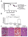
Anti-ER-TR7 inhibits tolerance induction. Tolerant mice received one dose of anti-ER-TR7 as follows: 100 μg d-6 (n=1), 200 μg d-6 (n=2), 50 μg d+1 (n=2), 100 μg d+1 (n=3), or 200 μg d+1 (n=2), relative to transplantation. An additional cohort of tolerant mice received one dose of rat IgG2a (rIgG2a) as follows: 200 μg d-6 (n=2), 200 μg d+1 (n=2), relative to transplant. Recipients received BALB/c heterotopic cardiac allografts on d0. Graft function monitored and recipients euthanized at time of transplant rejection or d25, and graft survival recorded (A). Grafts analyzed by H&E staining and PR score determined (B). (C) Black arrow indicates area of monocytic infiltrate, blue arrow indicates graft vascular occlusion in representative H&E staining images (600x, C). Data represented as percent survival (A) or mean ± SEM (B). n = 4–10 mice/group. * p < 0.05, ** p < 0.005, *** p < 0.0005.

References

    1. Katakai T, Hara T, Lee JH, Gonda H, Sugai M, Shimizu A. A novel reticular stromal structure in lymph node cortex: an immuno-platform for interactions among dendritic cells, T cells and B cells. Int Immunol. 2004;16 (8):1133. - PubMed
    1. Girard JP, Moussion C, Forster R. HEVs, lymphatics and homeostatic immune cell trafficking in lymph nodes. Nat Rev Immunol. 2012;12 (11):762. - PubMed
    1. Burrell BE, Ding Y, Nakayama Y, et al. Tolerance and lymphoid organ structure and function. Front Immunol. 2012;2:64. - PMC - PubMed
    1. Burrell BE, Bromberg JS. Fates of CD4+ T cells in a tolerant environment depend on timing and place of antigen exposure. Am J Transplant. 2012;12 (3):576. - PMC - PubMed
    1. Nakayama Y, Bromberg JS. Lymphotoxin-beta receptor blockade induces inflammation and fibrosis in tolerized cardiac allografts. Am J Transplant. 2012;12 (9):2322. - PMC - PubMed

Publication types

Substances