. 2015 Jun;9(6):1155-68.
doi: 10.1016/j.molonc.2015.02.007. Epub 2015 Feb 21.

Disulfiram (DSF) acts as a copper ionophore to induce copper-dependent oxidative stress and mediate anti-tumor efficacy in inflammatory breast cancer

Affiliations

Disulfiram (DSF) acts as a copper ionophore to induce copper-dependent oxidative stress and mediate anti-tumor efficacy in inflammatory breast cancer

Jennifer L Allensworth et al. Mol Oncol. 2015 Jun.

Abstract

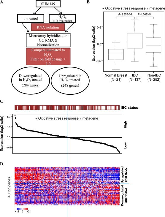
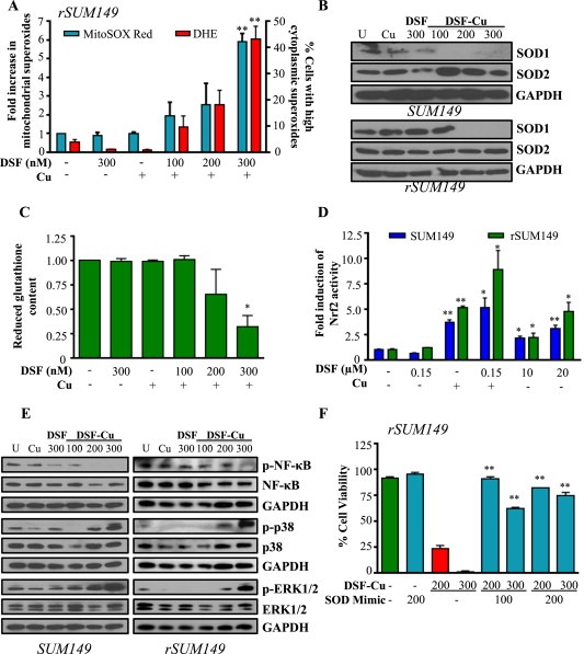
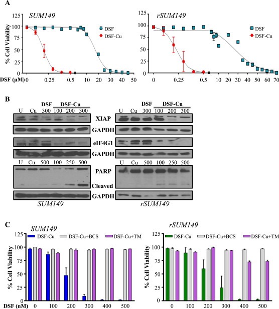
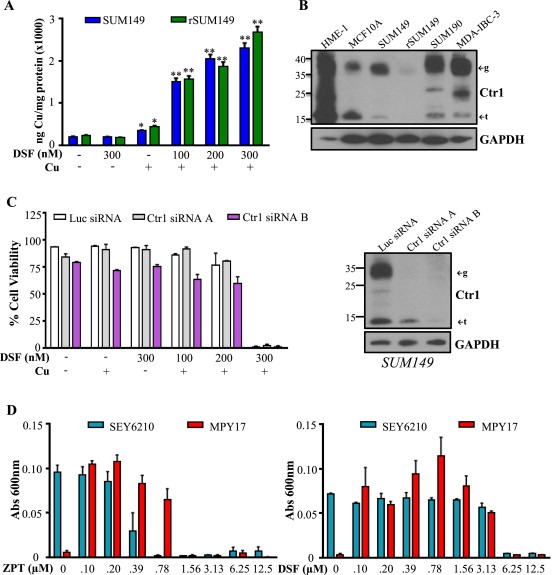
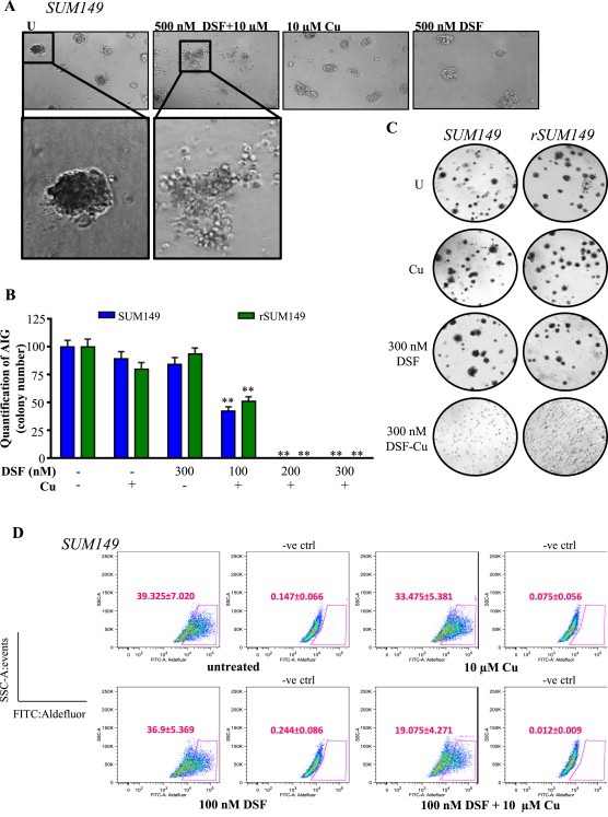
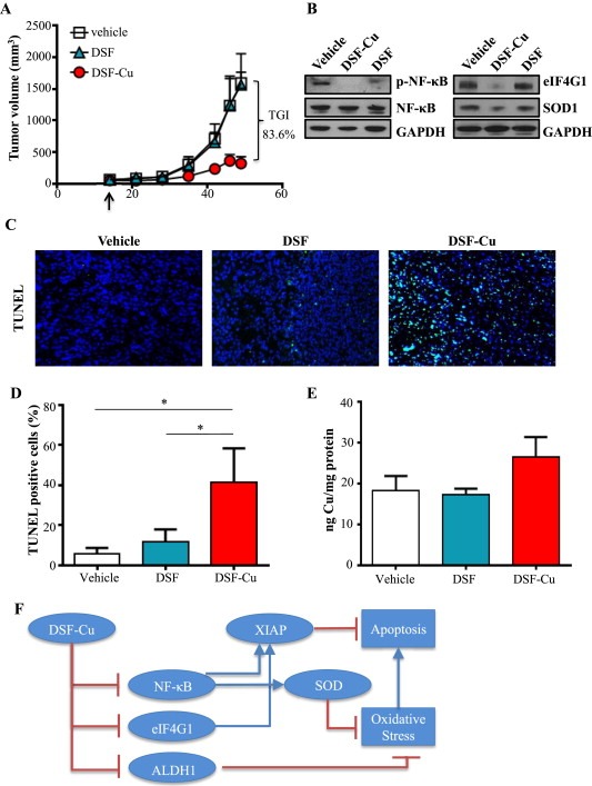
Cancer cells often have increased levels of reactive oxygen species (ROS); however, acquisition of redox adaptive mechanisms allows for evasion of ROS-mediated death. Inflammatory breast cancer (IBC) is a distinct, advanced BC subtype characterized by high rates of residual disease and recurrence despite advances in multimodality treatment. Using a cellular model of IBC, we identified an oxidative stress response (OSR) signature in surviving IBC cells after administration of an acute dose of an ROS inducer. Metagene analysis of patient samples revealed significantly higher OSR scores in IBC tumor samples compared to normal or non-IBC tissues, which may contribute to the poor response of IBC tumors to common treatment strategies, which often rely heavily on ROS induction. To combat this adaptation, we utilized a potent redox modulator, the FDA-approved small molecule Disulfiram (DSF), alone and in combination with copper. DSF forms a complex with copper (DSF-Cu) increasing intracellular copper concentration both in vitro and in vivo, bypassing the need for membrane transporters. DSF-Cu antagonized NFκB signaling, aldehyde dehydrogenase activity and antioxidant levels, inducing oxidative stress-mediated apoptosis in multiple IBC cellular models. In vivo, DSF-Cu significantly inhibited tumor growth without significant toxicity, causing apoptosis only in tumor cells. These results indicate that IBC tumors are highly redox adapted, which may render them resistant to ROS-inducing therapies. DSF, through redox modulation, may be a useful approach to enhance chemo- and/or radio-sensitivity for advanced BC subtypes where therapeutic resistance is an impediment to durable responses to current standard of care.

Keywords: ALDH; Antioxidant; Apoptosis; NFκB; SUM149; XIAP.


Figures

Figure 1
Figure 1
Oxidative stress response signature in IBC clinical samples. A, Schematic of development of oxidative stress response (OSR) metagene in SUM149 cells, comparing untreated to H2O2 administration. B, Expression values of the “oxidative stress response” metagene set generated from H2O2‐treated and untreated SUM149 cells; values are reported as a box plot according to the type of samples (normal breast, IBC, non‐IBC). p‐values are indicated (t‐test). C, Classification of 389 breast cancer samples from left to right based upon decreasing metagene value; the IBC/non‐IBC type is shown above the curve: red for IBC, white for non‐IBC. D, Heatmap showing expression values in BC samples from C for the top 40 genes most differentially expressed. Genes ordered from top to bottom according to decreasing log2‐ratio. Each row represents a gene and each column a sample. Expression levels are depicted according to the color scale at the bottom left, and color saturation represents the magnitude of deviation from the median.
Figure 2
Figure 2
DSF‐Cu reduces cellular antioxidant capacity to induce ROS and activate redox signaling. SUM149, rSUM149 cells treated with DSF, DSF + Cu (100–300 nM, 10 μM), Cu alone (10 μM). A, Fold induction of mitochondrial superoxides (teal bars) and percentage of cells with high cytoplasmic superoxides (red bars) measured by flow cytometry. B, Immunoblot analysis of SOD1/2. C, Reduced glutathione content relative to untreated (rSUM149 shown). D, Fold induction of Nrf2 activity measured by ARE‐responsive luciferase activity. E, Immunoblot analysis of indicated proteins in treated cells at 4 h time point. F, Effect of SOD mimetic (MnTnHexyl‐2‐Pyp5+, 100–200 μM) on viability measured by trypan blue exclusion assay (rSUM149 shown). *p < 0.05, **p < 0.005 in all panels. GAPDH and respective total proteins as loading controls.
Figure 3
Figure 3
DSF induces Cu‐dependent apoptosis. A, Dose‐dependent measurement of viability in cells treated with DSF, Cu (10 μM), or DSF‐Cu. B, Immunoblot analysis of apoptotic pathway proteins. GAPDH as loading control. C, Viability in the presence of Cu chelators bathocuproine disulphonate (BCS, 100 μM, gray bars) or tetrathiomolybdate (TM, 10 μM, purple bars) measured by trypan blue exclusion assay.
Figure 4
Figure 4
DSF acts as an ionophore to induce Ctr1‐independent Cu uptake. A, Cu content (ng) normalized to protein (mg) in SUM149 (blue bars) and rSUM149 (green bars) treated with DSF alone or in combination with Cu measured by ICP‐HRMS. *p < 0.05, **p < 0.005. B, Ctr1 expression in normal and IBC cell lines. ←g indicates glycosylated form, ←t indicates truncated form. GAPDH as loading control. C, Left: Viability of SUM149 cells treated with Ctr1‐targeting siRNAs (gray and purple bars) or control luciferase‐targeting siRNA (white bars) following DSF (300 nM), Cu (10 μM), or DSF‐Cu (100–300 nM, 10 μM) treatment for 24 h. Right: Immunoblot analysis of SUM149 cells treated with Ctr1‐targeting siRNAs (A and B) or control luciferase‐targeting siRNA. D, Growth of SEY6210 (blue bars) and Ctr1/3‐deficient MPY17 (red bars) S. cerevisiae cells in YPEG media with addition of ZPT (left) or DSF (right) measured by absorbance at 600 nm.
Figure 5
Figure 5
DSF‐Cu inhibits AIG and ALDH activity of IBC cells. A, Representative images of SUM149 mammospheres treated with indicated concentrations. Magnification: 10× inset: 20×. B, Quantification of AIG assay (by colony number) relative to untreated in SUM149 (blue bars) and rSUM149 (green bars) cells treated with DSF, Cu (10 μM), or DSF‐Cu complex. C, Representative AIG images of cells treated as indicated. D, Representative dot‐plots of ALDH1 activity. Cells were incubated with ALDEFLUOR substrate (BAAA), and the specific inhibitor of ALDH1, DEAB, was used to establish the baseline fluorescence and define ALDEFLUOR‐positivity (gated population). DEAB‐treated plots are labeled as –ve ctrl. Mean ± SEM of four independent experiments. Inset, Labeling of X and Y axes.
Figure 6
Figure 6
DSF‐Cu inhibits tumor growth in an in vivo model of IBC. A, Tumor volumes (measured V = (L × W2)/2) of mice with SUM149 subcutaneous flank tumors treated with vehicle, DSF, or DSF‐Cu. B, Representative immunoblot analysis of indicated proteins in tumor lysates from mice treated with vehicle, DSF, or DSF‐Cu. GAPDH as loading control. C, Representative images of tumor tissue from mice treated with vehicle, DSF, or DSF‐Cu with TUNEL staining. Magnification: 40×. DAPI (nuclei): blue; TUNEL (apoptotic cells): green. D, Quantification of TUNEL positive cells (from C). Mean ± SEM % TUNEL positive/total number of cells, *p < 0.05. E, Cu content of excised tumors (ng) measured by ICP‐HRMS relative to protein (mg). F, Schematic representation of DSF‐Cu mechanisms of action. DSF‐Cu complex acts as a pro‐oxidant, induces ROS‐mediated cancer cell death by inhibiting NFκB, which attenuates NFκB‐dependent antioxidant and anti‐apoptotic gene expression. DSF‐Cu inhibits ALDH1, which has been implicated in protection from ROS. DSF‐Cu also inhibits the potent anti‐apoptotic protein, XIAP, and translation initiation factor eIF4G1 (which can enhance XIAP translation during cell stress), promoting apoptosis.

References

    1. Abramoff, M.D. , Magalhaes, P.J. , Ram, S.J. , 2004. Image processing with ImageJ. Biophotonics Int. 11, 36–42.
    1. Aird, K.M. , Allensworth, J.L. , Batinic-Haberle, I. , Lyerly, H.K. , Dewhirst, M.W. , Devi, G.R. , 2012. ErbB1/2 tyrosine kinase inhibitor mediates oxidative stress-induced apoptosis in inflammatory breast cancer cells. Breast Cancer Res. Treat. 132, 109–119. - PMC - PubMed
    1. Aird, K.M. , Ding, X. , Baras, A. , Wei, J. , Morse, M.A. , Clay, T. , Lyerly, H.K. , Devi, G.R. , 2008. Trastuzumab signaling in ErbB2-overexpressing inflammatory breast cancer correlates with X-linked inhibitor of apoptosis protein expression. Mol. Cancer Ther. 7, 38–47. - PubMed
    1. Aird, K.M. , Ghanayem, R.B. , Peplinski, S. , Lyerly, H.K. , Devi, G.R. , 2010. X-linked inhibitor of apoptosis protein inhibits apoptosis in inflammatory breast cancer cells with acquired resistance to an ErbB1/2 tyrosine kinase inhibitor. Mol. Cancer Ther. 9, 1432–1442. - PMC - PubMed
    1. Allensworth, J.L. , Aird, K.M. , Aldrich, A.J. , Batinic-Haberle, I. , Devi, G.R. , 2012. XIAP inhibition and generation of reactive oxygen species enhances TRAIL sensitivity in inflammatory breast cancer cells. Mol. Cancer Ther. 11, 1518–1527. - PubMed

Publication types

MeSH terms