. 2015 Mar 17;42(3):524-37.
doi: 10.1016/j.immuni.2015.02.009. Epub 2015 Mar 10.

CXCR3 chemokine receptor enables local CD8(+) T cell migration for the destruction of virus-infected cells

Affiliations

CXCR3 chemokine receptor enables local CD8(+) T cell migration for the destruction of virus-infected cells

Heather D Hickman et al. Immunity. .

Abstract

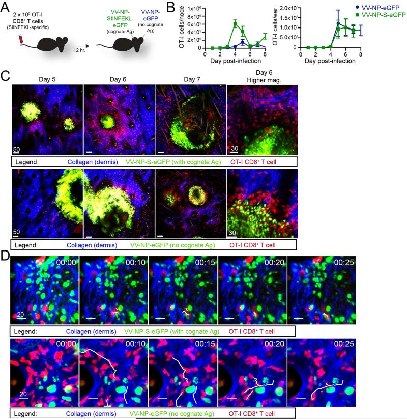
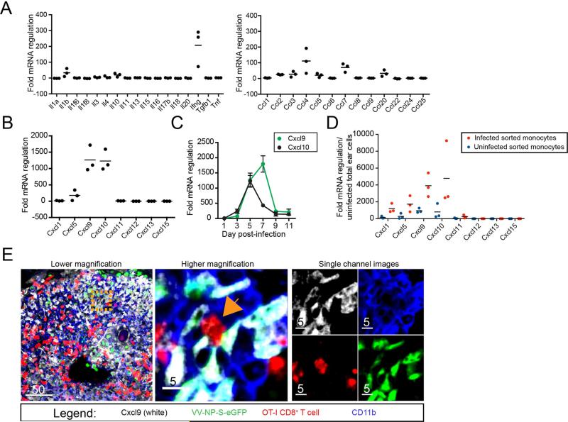
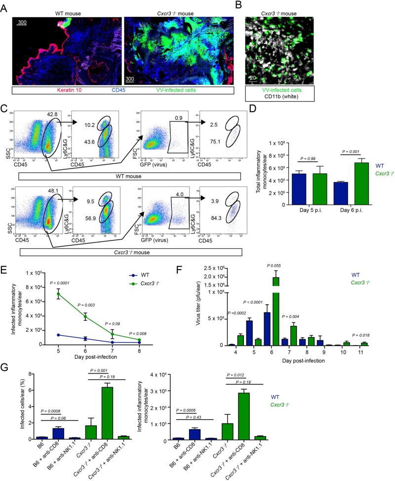
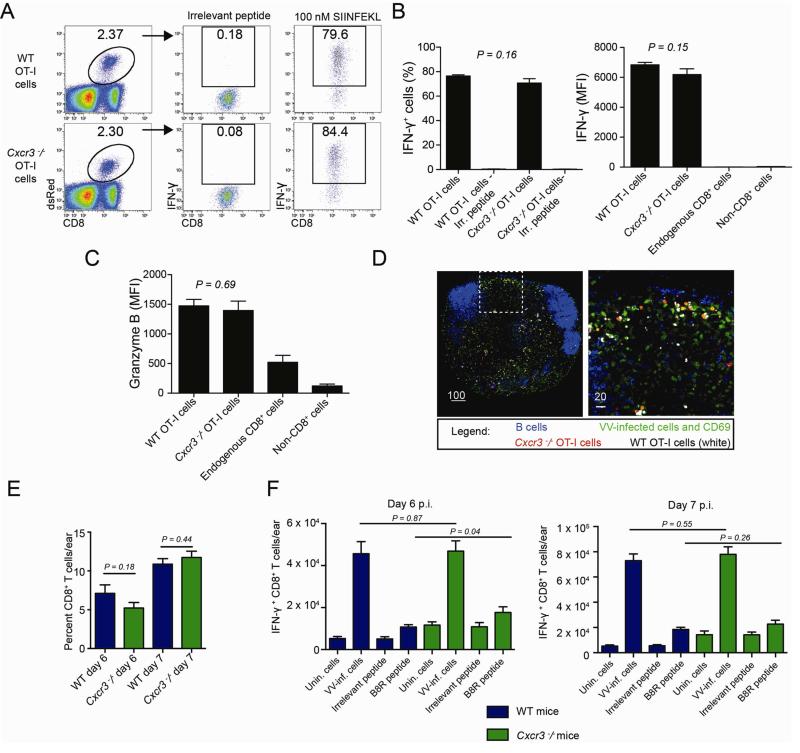
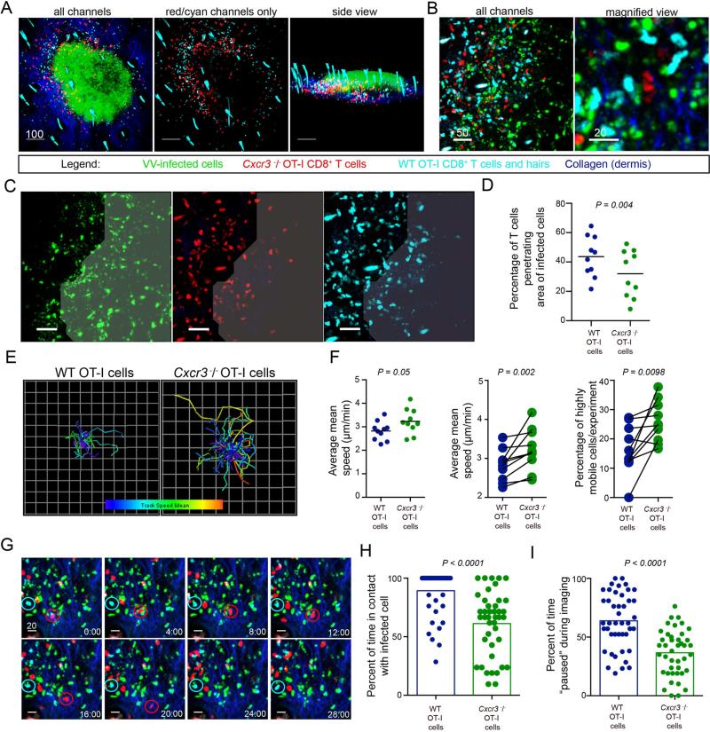
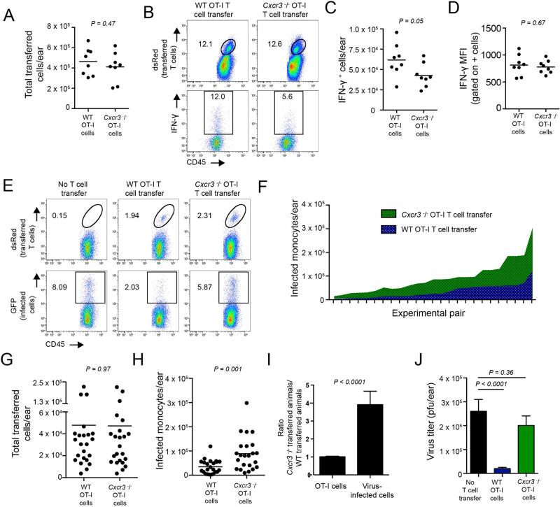
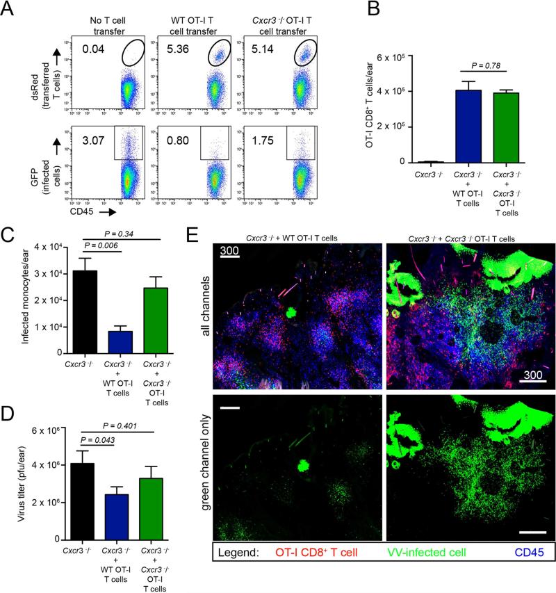
CD8(+) T cells play a critical role in limiting peripheral virus replication, yet how they locate virus-infected cells within tissues is unknown. Here, we have examined the environmental signals that CD8(+) T cells use to localize and eliminate virus-infected skin cells. Epicutaneous vaccinia virus (VV) infection, mimicking human smallpox vaccination, greatly increased expression of the CXCR3 chemokine receptor ligands CXCL9 and CXCL10 in VV-infected skin. Despite normal T cell numbers in the skin, Cxcr3(-/-) mice exhibited dramatically impaired CD8(+)-T-cell-dependent virus clearance. Intravital microscopy revealed that Cxcr3(-/-) T cells were markedly deficient in locating, engaging, and killing virus-infected cells. Further, transfer of wild-type CD8(+) T cells restored viral clearance in Cxcr3(-/-) animals. These findings demonstrate a function for CXCR3 in enhancing the ability of tissue-localized CD8(+) T cells to locate virus-infected cells and thereby exert anti-viral effector functions.


Figures

Figure 1
Figure 1. CD8+ T cells find virus-infected cells that don't express cognate antigen
A. Experimental design. 2.5 × 10⁵ OT-I T cells were transferred into wild-type (WT) animals prior to epicutaneous (ec.) infection in one ear with VV-NP-S-eGFP (with cognate Ag) and in the contralateral ear with NP-eGFP (no cognate Ag). B. OT-I cells per node (left) or ear (right) as determined by flow cytometric analysis. Infection with NP-S-eGFP (green); NP-e (blue). Time p.i. (days) = x axis. Error bars = SEM for 3-5 mice per group/timepoint. C. Maximum intensity projections (MIPs) of multiphoton microscopic (MPM) images of VV-infected ears taken at indicated day p.i. (top of images). Dermal collagen (blue); OT-I CD8+ T cells (red); virus-infected cells (green). Top panels = virus expressing SIINFEKL; bottom panels lacking SIINFEKL. Higher magnification images on day 6 p.i. (far right). D. Time-lapse MIP MPM images from day 5 p.i. with a contralateral infection as described in A. White lines = paths of selected CD8+ T cells during the 25 min. imaging period. Time (upper right hand) = min. All experiments were repeated 3 times with 3-5 mice/group. All scalebars = microns. See also Figure S1 and Movie S1.
Figure 2
Figure 2. CXCR3 ligands are upregulated in VV-infected skin
A. Fold regulation of mRNA in VV-infected ears 5 days p.i. compared to uninfected ears. Black lines = means. B. as in A. but showing α-chemokine mRNA changes. C. CXCL9 (green) and CXCL10 (blue) mRNA expression in infected skin at indicated day p.i. (x axis) D. Fold regulation of mRNA in uninfected (blue dots) or infected (red dots) inflammatory monocytes sorted from day 5 infected ears. E. Confocal images of frozen tissue sections 5 days p.i. with VV-NP-S-eGFP (green nuclear staining), CXCL9 (white), OT-I T cells (red) and CD11b (blue). Middle panel = higher magnification of orange dashed square from left panel. Orange arrow indicates an OT-I CD8+ T cell that is interacting with VV-infected CXCL9+ CD11b+ cells. Right panels = images of middle panel showing only one color (channel) for clarity. Scalebars = microns. All experiments were repeated 3 times. For mRNA data, dots = separate experiments with 2-5 mice/group. See also Figures S2 and S3.
Figure 3
Figure 3. Cxcr3−/− mice have elevated numbers of VV-infected inflammatory monocytes
A. Confocal montages of frozen sections of WT (left) or Cxcr3−/− ears 5 days p.i. (right). VV-infected cells (green), keratin 10 (red), CD45 (blue). B. Confocal image of Cxcr3−/− ear 5 days p.i. CD11b (white), VV-infected cells (green). C. Flow histograms from dissociated ear 5 days p.i. Top panels = wild-type (WT). Bottom panels = Cxcr3−/−. Middle left dot plots in both WT and Cxcr3−/− mice are gated on total cells. Middle right dot plots show infected (GFP+) cells. Far right dot plots are gated on infected cells and show the percentage of infected cells that are inflammatory monocytes (Ly6CG intermediate cells) and infected neutrophils (Ly6CG high cells). D. Total inflammatory monocytes on days 5-6 p.i. in WT (blue) or Cxcr3−/− (green) ears. E. Timecourse of infected inflammatory monocytes in WT (blue) or Cxcr3−/− (green) ears. F. Titer of infectious virus recovered per ear on day 5 or 6 p.i. WT (blue), Cxcr3−/− (green). G. Percentage and number of infected inflammatory monocytes in WT (blue) or Cxcr3−/− ears (green) day 6 p.i. Ab treatment group (performed prior to T cell entrance into the ear on days 4 and 5 p.i.) = x axis. Scalebars = microns. Error bars = SEM. Statistics = unpaired t tests. All experiments were repeated at least 3 times with 2-5 mice/group
Figure 4
Figure 4. Cxcr3−/− CD8+ T cells activate and home normally
A. Dot plots from lymph node cells 2 days p.i. with VV-NP-S-eGFP. Mice were given 2.5 × 10⁵ WT or Cxcr3−/− OT-I CD8+ T cells prior to infection. Gates show percentage of IFN-γ+ cells after restimulation with indicated peptide (top). B. Percentage (left) and MFI (right) of IFN-γ+ cells after restimulation. Endogenous CD8+ cells (all non-transferred CD8+ T cells in the lymph node) are shown for comparison. C. MFI of granzyme B. D. Frozen lymph node section 8 hr. p.i. with VV (green), B cells (blue), WT (white) or Cxcr3−/− (red) OT-I CD8+ T cells. CD69 is also stained green to indicate activated cells. E. Number of endogenous CD8+ T cells per ear days 6-7 p.i. WT = blue lines, Cxcr3−/− = green lines. F. Number of IFN-γ+ cells per ear in WT (blue) or Cxcr3−/− (green) mice on days 6 (left panel) or 7 (right panel) pi. Scalebars = microns. Error bars = SEM. Statistics = unpaired t tests. All experiments were repeated at least 3 times with 4-6 mice/group.
Figure 5
Figure 5. Cxcr3−/− CD8+ T cells have reduced entry into areas of infection and fail to efficiently contact virus-infected cells
A. MPM images of skin 5 days p.i. with VV (green), dermis (blue), WT (cyan) or Cxcr3−/− (red) OT-I CD8+ T cells. Note that autofluorescent hairs also appear cyan. B. MPM images from an area with many infected inflammatory monocytes (green). C. MPM images showing distribution of WT (cyan) or Cxcr3−/− (red) OT-I CD8+ T cells within a densely infected area (indicated by shading). For clarity, each color is shown independently. D. Percentage of WT (blue) or Cxcr3−/− (green) OT-I CD8+ T cells within densely infected areas. Dots represent individual microscopic fields. Line = mean. E. Overlaid tracks of T cells from the same field during a 30 min. imaging period. Tracks are color-coded from slow (blue) to fast (red). F. Average mean speeds of WT (blue) or Cxcr3−/− (green) OT-I CD8+ T cells from same field in an imaging session (left panel, statistics = Mann Whitney test). Middle panel = WT or Cxcr3−/− OT-I T cells in same field connected with a line (statistics = Wilcoxon test). Right panel shows percentage of cells moving > 2 SDs from mean speed of WT OT-I CD8+ T cells with lines indicating pairs in same imaging field (statistics = Wilcoxon test). G. Still frame MPM images of WT (cyan) or Cxcr3−/− (red) OT-I T cells over 30 min.. A WT cell (cyan circle) and a Cxcr3−/− cell (red circle) is followed over time. H. (Left) Percent of time WT (blue) and Cxcr3−/− (green) cells are contacting infected cells during 1 hr. imaging period. (Right) Percent of time paused on infected cell. Results were combined from two individual experiments and repeated twice. Time = min. Scalebars = microns. Error bars = SEM. All experiments except H were repeated at least 3 times with 4-6 mice/group. See also Figure S4 and Movies S2-S7.
Figure 6
Figure 6. Cxcr3−/− CD8+ T cells do not kill infected cells as efficiently as WT CD8+T cells
A. Number of WT or Cxcr3−/− OT-I CD8+ T cells on day 5 p.i. after adoptive transfer into WT animals. Dots = individual ears. B. Histograms gated on percentage of transferred T cells (dsRed+, top panels) or on IFN-γ+ transferred T cells (bottom panels). WT OT-I cells (left), Cxcr3−/− OT-I cells (right). C. Number of IFN-γ+ cells/ear on day 5 p.i. D. Mean fluorescent intensity (MFI) of IFN-γ signal from IFN-γ+ population of transferred cells. E. Percentage of transferred cells (top panels) recovered in Cd8−/− ears after transfer of 2.5 × 10⁵ OT-I cells. No transfer (left panels), WT OT-I cell transfer (middle panels) and Cxcr3−/− OT-I transfer (right panels). F. Number of infected cells in Cd8−/− ears with titrated numbers (from 2.5 × 10³ – 7.5 × 10⁵) of transferred WT (blue) or Cxcr3−/− T cells (green). X axis shows pairs of CD8−/− mice in which equivalent numbers of transferred OT-I T cells were recovered. G. Number of T cells recovered in Cd8−/− mice in experiment shown in F. H. Number of infected monocytes per ear from experiment F. I. Ratios from mice in F. (Left bar) Number of transferred Cxcr3−/− OT-I cells recovered divided by the number of WT OT-I cells recovered. (Right bar) Number of infected cells under either condition. Statistics in A-C= two-tailed unpaired t test. Statistics in F= two-tailed paired t test. Error bars = SEM. Experiments A-C were performed 3 times with 3-4 mice/group. Experiments F-G were performed in 5 independent experiments with 2-3 mice/group.
Figure 7
Figure 7. Transfer of WT OT-I CD8+ T cells into Cxcr3−/− animals restores viral control
A. Dot plots from Cxcr3−/− mice with no T cell transfer (left), or with 2.5 × 10⁵ WT (middle) or Cxcr3−/− (right) OT-I T cells on 5 days p.i. B. Number of adoptively transferred WT or Cxcr3−/− OT-I CD8+ T cells recovered per ear in Cxcr3−/− animals on day 5 p.i. C. Number of virus-infected cells per ear in WT or Cxcr3−/− OT-I CD8+ T cell adoptively-transferred Cxcr3−/− animals. D. Viral titers from C. E. Confocal montage images of Cxcr3−/− ears 5 days p.i. with VV (green) with transfer of WT or Cxcr3−/− OT-I T cells (red) before infection. CD45+ cells = blue. For clarity, the green channel only is shown in the bottom panels. Statistics= unpaired two-tailed t test. Error bars = SEM. Scalebars = μms.

Comment in

References

    1. Cole KE, Strick CA, Paradis TJ, Ogborne KT, Loetscher M, Gladue RP, Lin W, Boyd JG, Moser B, Wood DE, et al. Interferon-inducible T cell alpha chemoattractant (I-TAC): a novel non-ELR CXC chemokine with potent activity on activated T cells through selective high affinity binding to CXCR3. J Exp Med. 1998;187:2009–2021. - PMC - PubMed
    1. Friedl P, Weigelin B. Interstitial leukocyte migration and immune function. Nat Immunol. 2008;9:960–969. - PubMed
    1. Gordon SN, Cecchinato V, Andresen V, Heraud JM, Hryniewicz A, Parks RW, Venzon D, Chung HK, Karpova T, McNally J, et al. Smallpox vaccine safety is dependent on T cells and not B cells. J Infect Dis. 2011;203:1043–1053. - PMC - PubMed
    1. Groom JR, Luster AD. CXCR3 in T cell function. Experimental cell research. 2011a;317:620–631. - PMC - PubMed
    1. Groom JR, Luster AD. CXCR3 ligands: redundant, collaborative and antagonistic functions. Immunol Cell Biol. 2011b;89:207–215. - PMC - PubMed

Publication types

MeSH terms