. 2015 May 15;75(10):1972-82.
doi: 10.1158/0008-5472.CAN-14-2761. Epub 2015 Mar 13.

Paracrine WNT5A Signaling Inhibits Expansion of Tumor-Initiating Cells

Affiliations

Paracrine WNT5A Signaling Inhibits Expansion of Tumor-Initiating Cells

Nicholas Borcherding et al. Cancer Res. .

Abstract

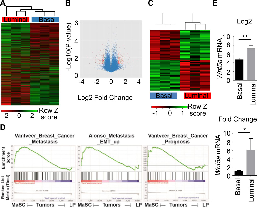
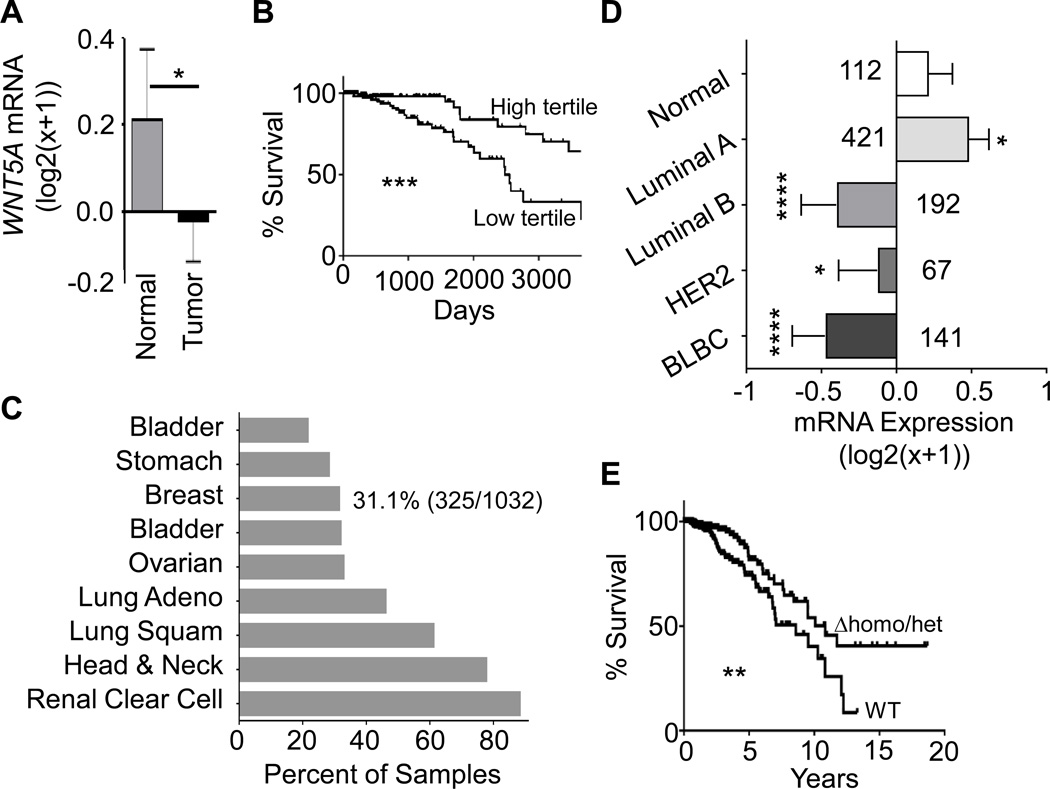
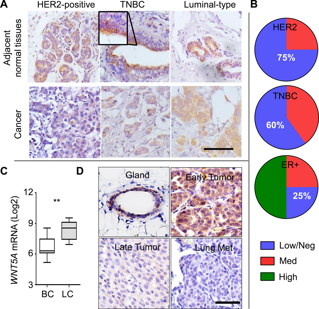
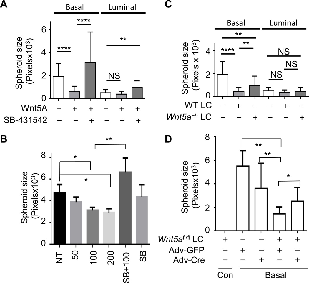
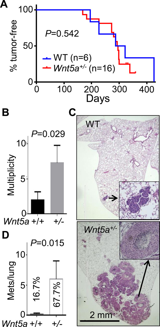
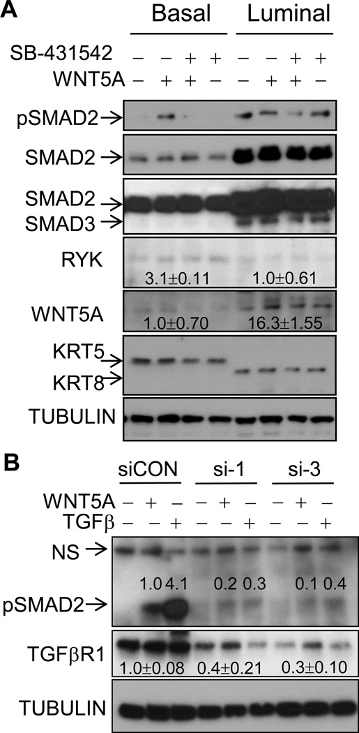
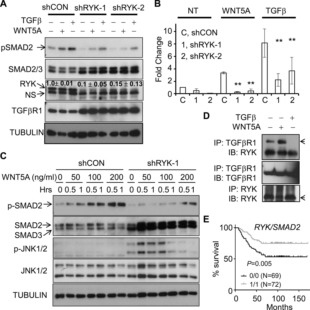
It is not well understood how paracrine communication between basal and luminal cell populations in the mammary gland affects tumorigenesis. During ErbB2-induced mammary tumorigenesis, enriched mammary stem cells that represent a subpopulation of basal cells exhibit enhanced tumorigenic capacity compared with the corresponding luminal progenitors. Transcript profiling of tumors derived from basal and luminal tumor-initiating cells (TIC) revealed preferential loss of the noncanonical Wnt ligand WNT5A in basal TIC-derived tumors. Heterozygous loss of WNT5A was correlated with shorter survival of breast cancer patients. In a mouse model of ErbB2-induced breast cancer, Wnt5a heterozygosity promoted tumor multiplicity and pulmonary metastasis. As a TGFβ substrate, luminal cell-produced WNT5A induced a feed-forward loop to activate SMAD2 in a RYK and TGFβR1-dependent manner to limit the expansion of basal TIC in a paracrine fashion, a potential explanation for the suppressive effect of WNT5A in mammary tumorigenesis. Our results identify the WNT5A/RYK module as a spatial regulator of the TGFβ-SMAD signaling pathway in the context of mammary gland development and carcinogenesis, offering a new perspective on tumor suppression provided by basal-luminal cross-talk in normal mammary tissue.


Conflict of interest statement

Conflict of Interest

All authors possess no conflicts of interest.

Figures

Figure 1
Figure 1. Transcriptome comparison between basal-TIC and luminal-TIC tumors
A. Heatmap for unsupervised clustering of gene profiles from paired tumors derived from basal TICs (Basal, red) and luminal TICs (Luminal, blue). The whole transcriptomes were clustered using R gplots. n=3/group. B. Volcano plot showing differentially-expressed between luminal- and basal-TIC tumors, with P-value < 0.05 and log2 fold-change > 1.58 (linear 3-fold change) highlighted in red. C. 55 genes were identified to fit the criteria in B, with exclusive expression in basal TIC- and luminal TIC-derived tumors. D. Gene Set Enrichment Analysis (GSEA) was used to analyze the difference of biological signaling pathways between basal-TIC or luminal-TIC formed tumors based transcriptome data from A. Basal TIC tumors upregulate signaling pathways related to breast cancer metastasis and bad prognosis. Nominal P values < 0.001 for pathways mentioned. E. WNT5A expression in mouse basal-TIC and luminal-TIC tumors. Both Log2 values (Top) and linear fold-change (Bottom) are shown, P = 0.0076, n=3).
Figure 2
Figure 2. Heterozygous loss of WNT5A is common in cancer
A. WNT5A expression in TCGA Illumina HiSeq level 3 breast-cancer dataset by sample type (P = 0.0154, n= 112 for normal tissues and n= 1040 for invasive cancer). B. Kaplan-Meier curve to show the overall survival between the highest WNT5A-expressing samples (n=126) and the lowest WNT5A-expressing samples (n=125) among the Agilent G4502A level 3 microarray TCGA BRCA dataset. ***: P = 0.0003. C. Copy number variations (CNV) from different TCGA datasets were analyzed using UCSC cancer genome browser and plotted as percentages of specimens with any loss of WNT5A alleles. D. WNT5A expression in BRCA TCGA Illumina HiSeq level 3 by PAM50 molecular subtype. P values comparing Normal samples (n=60) to Luminal A (n=211, P = 0.0142), Luminal B (n=128, P < 0.0001), HER2 (n=58, P = 0.0389), and basal-like breast cancer (BLBC, n=78, P < 0.0001). E. Kaplan-Meier curve to show the overall survival between WNT5A bialleles (WT, n=519) and either homozygous or heterozygous deletion of WNT5A alleles (Δhomo/het, n=249) among Illumina HiSeq RNAseq level 3 BRCA data. **: P = 0.0098.
Figure 3
Figure 3. WNT5A protein is lost in more aggressive types of breast cancer
A. Representative WNT5A immunohistochemistry (IHC) staining for triple negative breast cancer (TNBC), HER2+, or estrogen receptor (ER)+ breast cancer specimens (bottom panels) with adjacent normal mammary tissues (top panels) (bar = 100 µm). B. Quantification of WNT5A staining in HER2-positive (n=4), TNBC (n=5), and ER-positive luminal type (n=4) patient tissue samples. C. Microarray dataset GSE37223 deposited in GEO Datasets in Pubmed Central was downloaded and analyzed for the expression of WNT5A in CD49f+EPCAM human basal mammary epithelial cells and CD49fEPCAM+ luminal epithelial cells (P = 0.0093, n=7 for each population). D. Representative WNT5A IHC staining from MMTV-ErbB2 mammary glands and tumors. Early tumors were collected at 5–6 months of age when tumors were within 0.5 cm; whereas late tumors (2.5 cm) and paired lung metastases were collected at 7–8 months of age (bar = 50 µm). n=3 for each type.
Figure 4
Figure 4. WNT5A suppresses the growth of basal TIC via a paracrine manner
A. Basal and luminal TICs were purified from pre-neoplastic mammary glands of 5-month old MMTV-ErbB2 female mice and seeded onto matrigel-coated chambers, either mock-treated, or treated with 100 ng/ml of WNT5A alone, or in combination with 5 µM of SB-431542. B. Basal TICs were purified and seeded onto matrigel-coated chambers, either non-treated (NT), treated with different doses of WNT5A (50, 100, or 200 ng/ml), or 100ng/ml WNT5A + 5 µM SB-431542 (SB+100), or 5 µM SB-431542 (SB). C. Basal and luminal TICs were purified and seeded onto matrigel, either alone or co-cultured with purified mature luminal cells (LC) from either WT or WNT5A+/− female mice at 1:5 ratio. D. Basal TICs (basal) and mature luminal cells from WNT5Afl/fl females (WNT5Afl/fl LC) were co-seeded onto matrigel at 1:5 ratio; 24 hrs later, cells were either treated with adenovirus encoding GFP (Adv-GFP) or Cre DNA recombinase (Adv-Cre). Basal TICs alone were also treated with Adv-GFP or Adv-Cre. WNT5Afl/fl LC did not form spheroids under these conditions (WNT5Afl/fl LC, first column). A–D. After 3 weeks, spheroids were photographed and their size in pixels was quantitated with Image J ((*<0.05, ** P ≤ 0.0042, **** P < 0.0001). Mean ± s.e.m., n=10–27 spheroids from two independent experiments).
Figure 5
Figure 5. WNT5A inhibits tumor multiplicity and metastasis in ErbB2-induced mammary cancer
MMTV-ErbB2 transgenic mice were crossed with WNT5A+/− to produce ErbB2/WT and ErbB2/WNT5A+/− mice. Tumor onset days (A), multiplicity (B), as well as lung metastasis assessed by hematoxylin & eosin staining of paraffin-embedded lung sections (representative images in C and summarized in D) were assessed. % in C indicates the percentage of animals with lung metastasis. Numbers of metastatic nodules were plotted and P values indicated. A. n=6 for ErbB2/WT and n=16 for ErbB2/WNT5A+/−; B, D: n=6 for each group.
Figure 6
Figure 6. WNT5A induces the phosphorylation of SMAD2 in a TGFβ receptor-dependent fashion
A. Basal and luminal mammary epithelial cells were purified from pre-neoplastic mammary gland of 5-month-old ErbB2/WNT5A+/− mice and cultured in 24 well plates for 72 hrs. Cells were either mock-treated, treated with WNT5A (100ng/ml), WNT5A+SB-431542 (5 µM), or SB-431542 alone for 1 hr. B. MCF10A cells were transiently transfected with control siRNA (siCON), or two independent siRNAs to TGFβR1 (si-1, si-3). 96 hrs later, cells were either treated with WNT5A (100ng/ml) or TGFβ (5 ng/ml) for 1 hr. For A–B, cell lysates were collected, separated by SDS-PAGE and immunoblotted with the indicated antibodies (n=2–3). Band densitometry was measured by Image J.
Figure 7
Figure 7. RYK is the co-receptor for WNT5A-induced activation of SMAD2
A–B. MCF10A cells were infected with lentiviral particles encoding control luciferase shRNA (shCON), or two independent RYK shRNAs with co-expression of GFP. GFP-positive cells were collected by flow cytometry. Parental MCF10A (data not shown here), MCF10A cells with control shRNA, or cells with RYK shRNA1 or shRNA2 were cultured and either left untreated (NT) or treated with WNT5A (100 ng/ml) or TGFβ (5 ng/ml). Cell lysates were collected, separated by SDS-PAGE and immunoblotted with the indicated antibodies. Representative images were shown in A. and quantitated in B. (P < 0.01, n=3). C. MCF10A cells with control shRNA, or cells with RYK shRNA1 were treated with different amount of WNT5A as indicated for 0.5 or 1 hr. Cell lysates were collected, followed by immunoblotting with the indicated antibodies. D. MCF10A cells were treated with WNT5A (100ng/ml) or TGFβ1 (5 ng/ml) for 1 hr. Cells were lysed and lysates were immunoprecipitated with anti-RYK and anti-TGFβR1 antibodies. Immunocomplex was resolved by SDS-PAGE, followed by immunoblotting with the indicated antibodies. E. Affymetrix GEO dataset GSE2034 containing 255 human breast cancer specimens with supplemented clinical records was downloaded. 255 specimens in GSE2034 were separated into four groups based on the expression level of RYK and SMAD2. Low (assigned as 0) and high (assigned as 1) expression of RYK or SMAD2 were defined as expression values lower or higher than median values. N numbers and P value comparing 1/1 group VS. 0.0 group is indicated. The correlation between metastasis-free survival and RYK/SMAD2 expression was analyzed and Kaplan-Meier curve was graphed using Prism 6 software.

Similar articles

Cited by

References

    1. Taipale J, Beachy PA. The Hedgehog and Wnt signalling pathways in cancer. Nature. 2001;411(6835):349–354. - PubMed
    1. Habas R, Dawid IB, He X. Coactivation of Rac and Rho by Wnt/Frizzled signaling is required for vertebrate gastrulation. Genes Dev. 2003;17(2):295–309. - PMC - PubMed
    1. Wansleeben C, Meijlink F. The planar cell polarity pathway in vertebrate development. Developmental Dynamics. 2011;240(3):616–626. - PubMed
    1. Slusarski DC, Yang-Snyder J, Busa WB, Moon RT. Modulation of Embryonic Intracellular Ca2+Signaling byWnt-5A. Developmental Biology. 1997;182(1):114–120. - PubMed
    1. Westfall TA, Brimeyer R, Twedt J, Gladon J, Olberding A, Furutani-Seiki M, et al. Wnt-5/pipetail functions in vertebrate axis formation as a negative regulator of Wnt/β-catenin activity. J Cell Biol. 2003;162(5):889–898. - PMC - PubMed

Publication types

MeSH terms