. 2015 May;200(1):331-42.
doi: 10.1534/genetics.115.174920. Epub 2015 Mar 13.

A single gene causes an interspecific difference in pigmentation in Drosophila

Affiliations

A single gene causes an interspecific difference in pigmentation in Drosophila

Yasir H Ahmed-Braimah et al. Genetics. 2015 May.

Abstract

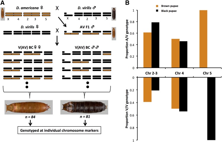
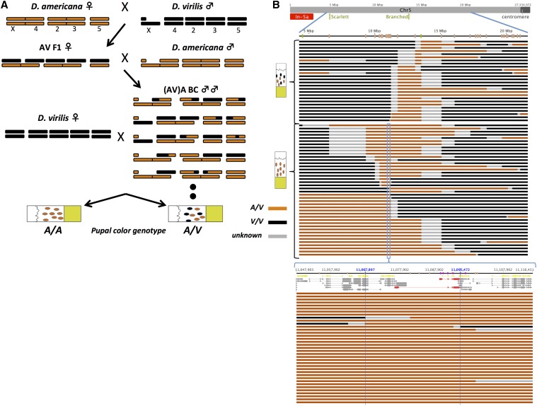
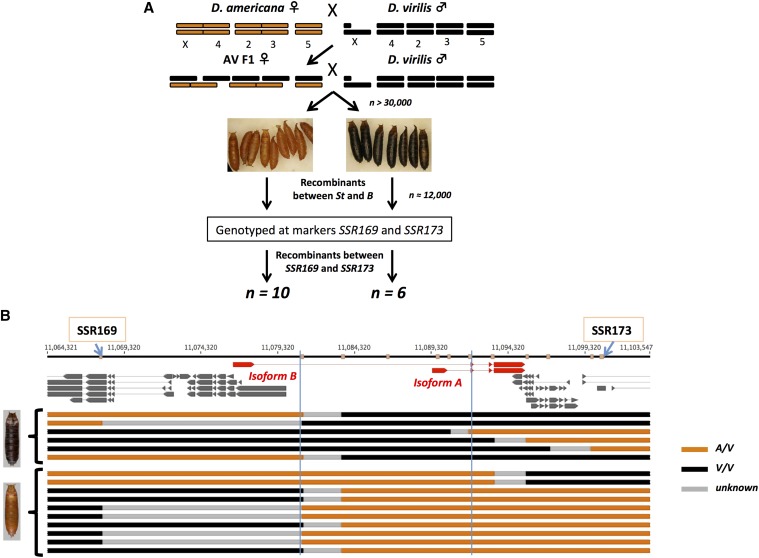
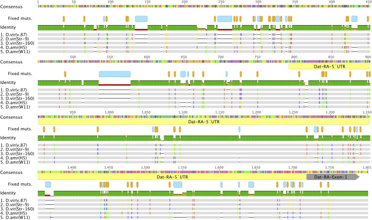
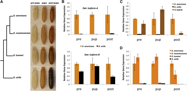
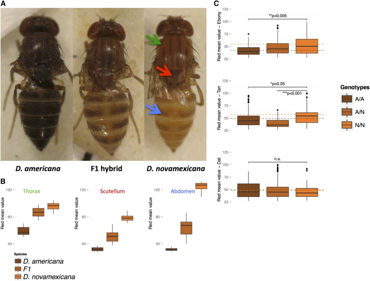
The genetic basis of species differences remains understudied. Studies in insects have contributed significantly to our understanding of morphological evolution. Pigmentation traits in particular have received a great deal of attention and several genes in the insect pigmentation pathway have been implicated in inter- and intraspecific differences. Nonetheless, much remains unknown about many of the genes in this pathway and their potential role in understudied taxa. Here we genetically analyze the puparium color difference between members of the virilis group of Drosophila. The puparium of Drosophila virilis is black, while those of D. americana, D. novamexicana, and D. lummei are brown. We used a series of backcross hybrid populations between D. americana and D. virilis to map the genomic interval responsible for the difference between this species pair. First, we show that the pupal case color difference is caused by a single Mendelizing factor, which we ultimately map to an ∼11-kb region on chromosome 5. The mapped interval includes only the first exon and regulatory region(s) of the dopamine N-acetyltransferase gene (Dat). This gene encodes an enzyme that is known to play a part in the insect pigmentation pathway. Second, we show that this gene is highly expressed at the onset of pupation in light brown taxa (D. americana and D. novamexicana) relative to D. virilis, but not in the dark brown D. lummei. Finally, we examine the role of Dat in adult pigmentation between D. americana (heavily melanized) and D. novamexicana (lightly melanized) and find no discernible effect of this gene in adults. Our results demonstrate that a single gene is entirely or almost entirely responsible for a morphological difference between species.

Keywords: genetics of species differences; morphological evolution; pigmentation.


Figures

Figure 1
Figure 1
(A) Crossing scheme to generate backcross pupae segregating for whole chromosomes. D. americana (A) and D. virilis (V) chromosomes are shown in brown and black, respectively. Female genotypes are shown on the left and male genotypes on the right. For each hybrid the genotype notation is abbreviated and the maternal allele is indicated first. The pupal color phenotype for pure species and hybrids is shown next to the pure species and F1 karyotypes. Eight possible genotypes (four female and four male) are illustrated for backcross hybrids. (B) Associations between chromosomal genotypes and pupal color phenotype among whole-chromosome backcross hybrids. The proportion of the two alternative whole-chromosome genotypes for red/brown and black backcross pupae is plotted for each autosomal linkage group. The sum of A/V and V/V genotypes for each phenotypic class within each chromosome should be 1.
Figure 2
Figure 2
(A) Crossing scheme to generate recombinant backcross hybrid males. Color scheme and notation of male and female genotypes is the same as in Figure 1A. The two possible pupal color phenotypes and the corresponding genotypes at the pupal color locus for the recombinant backcross males are indicated at the bottom. (B) Genotypes of recombinant backcross males along chromosome 5. The gray line at the top represents the entire length of chromosome 5, with the location of the two visible mutations indicated in green and inversion 5a indicated in red. Blue lines magnify the molecularly genotyped region, where the location of microsatellite markers is indicated in orange. Each horizontal line below the chromosome represents a single recombinant backcross male, with regions along the chromosome color coded according to inferred genotypes using microsatellite markers; brown, A/A; black, A/V; gray, unknown. Recombination breakpoints reside within gray regions. Recombinant males are grouped according to pupal color phenotype (shown on the left). The pupal color locus resides in a black region (A/V) among recombinant males who sire both black and light brown pupae (top group), or in a light brown region (A/A) among recombinant males who sire light brown pupae only (bottom group). Bottom: close-up of a 68.5-kb candidate region among recombinant backcross males who sired light brown pupae only. Top: chromosomal coordinates and markers (orange, Microsattelite; purple, SNP) along with the gene annotations from Flybase (yellow) and those inferred by Cufflinks (gray). GJ20215 is shown in red (top, isoform A; bottom, isoform B).
Figure 3
Figure 3
(A) Crossing scheme to enrich for recombinants in the candidate region. (B) Recombinant individuals recovered using the recombination enrichment strategy. Only Cufflinks assembled transcripts are shown and GJ20215 is highlighted in red. The recombinants recovered are grouped by pupal color phenotype, where black indicates D. virilis homozygous regions (V/V) and light brown are heterozygous regions. The two vertical blue lines indicate the mapped candidate interval.
Figure 4
Figure 4
Multiple sequence alignment of a representative 1.8-kb region. This alignment shows a portion of the ∼11-kb candidate region, which includes ∼800 bp upstream of the transcription start site of GJ20215 isoform A, the 5′-untranslated region (yellow), and the first exon (gray). Fixed differences between D. americana and D. virilis are indicated in orange (nucleotide substitutions) and blue (insertion/deletions) (produced with Geneious R8).
Figure 5
Figure 5
(A) Virilis group cladogram and pupae from the four virilis phylad members across the three developmentally defined stages used in RT–qPCR experiments. (B) Relative gene expression measures for the two isoforms in D. americana and D. virilis. Error bars represent standard error. All D. americana samples are normalized to 1 and considered the control sample. All comparisons are significant except for the “pup” sample of isoform B (P < 0.05). (C) Relative gene expression measures of GJ20215 isoform A in D. americana, D. virilis, and F1 hybrids. (c) Relative gene expression measures in all four species of the virilis group.
Figure 6
Figure 6
(A) Female D. americana, D. novamexicana, and F1 hybrids (14 days old). Arrows in the D. novamexicana image point to the adult landmarks used in quantifying emitted red values (arrow colors correspond to landmarks in B). (b) Distribution of mean emitted red values (represented by box plots) across a sample of D. americana, D. novamexicana, and F1 hybrids. (C) Box plot distribution of mean red values in the abdomen across genotypes for the three pigmentation genes surveyed. Red mean values are partitioned into the three genotypic classes recovered in the F6 population (n = 188). Dashed horizontal lines indicate the mean read value for A/A (brown) and N/N (orange) genotypes. Significant results from one-way ANOVA tests are indicated above each plot with asterisks and P-value, whereas the nonsignificant result is labeled n.s.

References

    1. Ahmed-Braimah Y. H., McAllister B. F., 2012. Rapid evolution of assortative fertilization between recently allopatric species of Drosophila. Int. J. Evol. Biol. 2012: 1–9. - PMC - PubMed
    1. Arnoult L., Su K. F. Y., Manoel D., Minervino C., Magriña J., et al. , 2013. Emergence and diversification of fly pigmentation through evolution of a gene regulatory module. Science 339: 1423–1426. - PubMed
    1. Bastide, H., A. Betancourt, V. Nolte, R. Tobler, P. Stöbe et al., 2013 A genome-wide, fine-scale map of natural pigmentation variation in Drosophila melanogaster. PLoS Genet. 9: e1003534. - PMC - PubMed
    1. Benson G., 1999. Tandem repeats finder: a program to analyze DNA sequences. Nuc. Ac. Res. 27: 573–580. - PMC - PubMed
    1. Blight W. C., Romano A., 1953. Notes on a breeding site of Drosophila americana near St-Louis, Missouri. Am. Nat. 87: 111–112.

Publication types

LinkOut - more resources